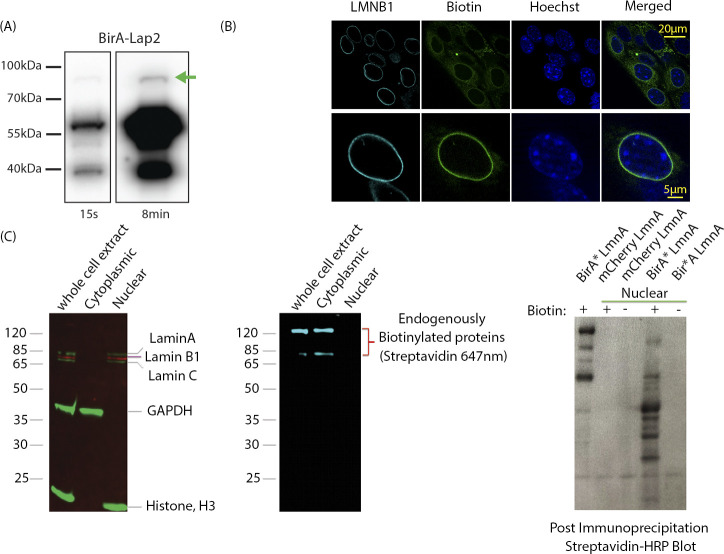
Figure S1.
Validation of BirA*-Lap2b expression and nuclear protein enrichment strategy. (A) Western blot showing relative levels of BirA-Lap2β levels at 2 different exposures (15 s, 8 min). BirA-Lap2β fusion protein is indicated by the green arrow and is expressed at 1–2% of endogenous Lap2β. (Antibody BD611000) (B) Prevalence of BirA-Lap2b-driven biotinylation in MEFs. Cells were stained for Lamin B1 (cyan), Bitoin (streptavidin-488, green), and DNA (Hoechst). >75% of cells display inner nuclear membrane enrichment of biotinylated proteins. Cells with low or no inner nuclear membrane enrichment still have biotinylation signal, but predominantly in the mitochondria (e.g., upper panel, upper left). Upper panel shows a field view of cells, lower panel a single cell. (C) Nuclear enrichment removes endogenously biotinylated proteins (middle panel) while retaining lamin components (left panel). In a protocol development strategy (right panel), BirA* tagged Lamin A extracts were subjected to detection with HRP-streptavidin with and without nuclear extraction.(and +/− Biotin). Without nuclear extraction, most detected proteins are the endogenously labelled mitochondrial proteins (right lane, biotin (+) and BirA* LmnA). After extraction, most proteins are new and specific to Lamin A marking (BirA* LmnA versus mCherry LmnA).

