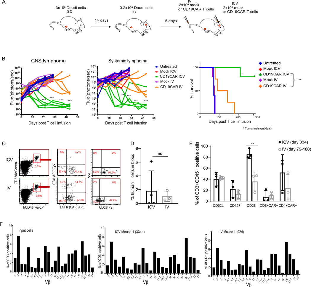
Figure 2. CD19-CAR T cells administered by ICV infusion can completely eradicate both CNS and systemic lymphoma.
(A) NSG mice were injected subcutaneously (SC) with 3×10⁶ EGFP+ ffluc+ Daudi lymphoma cells and 14 days later injected intracranially (IC) with 0.2×10⁶ EGFP+ ffluc+ Daudi lymphoma cells. Five days following IC tumor inoculation, 2×10⁶ mock-transduced T cells or CD19-CAR T cells were administered either ICV or IV. (B) Flux (photons/sec) was determined by measuring bioluminescence once weekly. N=5 mice per group. Linear mixed models were used to compare logarithm transformed flux of ICV- and IV-treated mice over time; **P<0.01, ***P<0.001. Survival of mice was analyzed by Log-rank test; **P<0.01. (C-E) At the time of euthanasia (day 334 for the ICV group and between days 79–180 for the IV group) blood was collected and analyzed for expression of human CD45, CD8, CD4, CAR (EGFR) and central memory receptors by flow cytometry. Representative data from two separate experiments are presented. Percentages (mean ±SD) of human T cells in the blood (D) and T cells expressing CD62L, CD127, CD28, and CAR+ CD4 and CD8 T cell subsets (E) are presented. ICV and IV were compared by Student’s t test. **P<0.01. (F) Human T cells harvested from mouse spleens 334 days post ICV and 180 days post IV CAR T cell infusion were re-stimulated with REM method containing OKT3, irradiated PBMC, and LCL for 14 days and their TCR repertoire was analyzed. Percent of TCR Vβ expression in CD3 positive cells is depicted.

