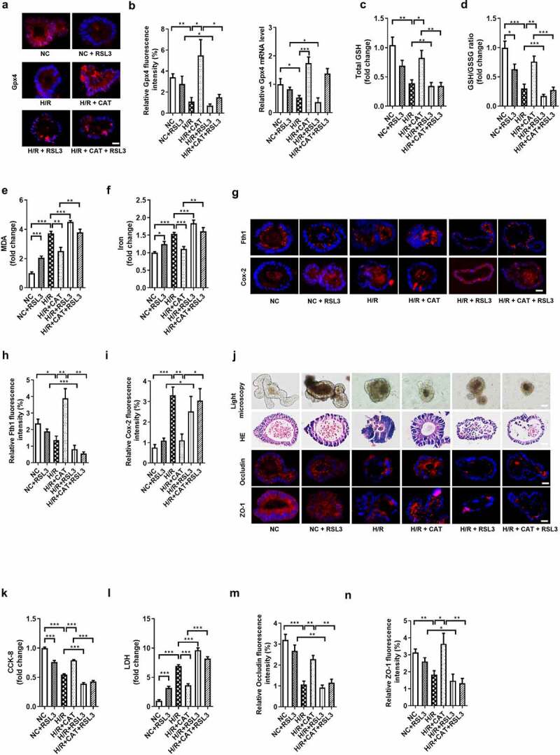
Figure 6.

CAT inhibits ferroptosis-dependent intestinal organoid H/R injury by promoting Gpx4 expression in vitro. (a)The protein levels of Gpx4 in the organoids, scale bar is 20 μm. (b) Relative fluorescence quantitative statistics of Gpx4 protein expression and relative Gpx4 mRNA level in the organoids. (c-d) Total GSH and the ratio of total GSH/GSSG in organoids. (e-f) MDA and Fe2+ levels in the organoids. (g) The protein levels of Fth1 and Cox-2, scale bar is 20 μm. (h-i) Relative fluorescence quantitative statistics of Fth1 and Cox-2 protein expression. (j) Ileum organoid morphology observed by light microscopy and HE staining and the expression of Occludin and ZO-1 in the organoids observed by immunofluorescence, scale bar is 20 μm. (k) Organoid viability measured by CCK-8. (l) Levels of LDH released into the medium. (m-n) Relative fluorescence quantitative statistics of Occludin and ZO-1 protein expression. The results are expressed as the mean ± SEM. n = 6. * p < .05, ** p < .01, *** p < .001 by one-way ANOVA (Tukey’s test)
