Figure 5.

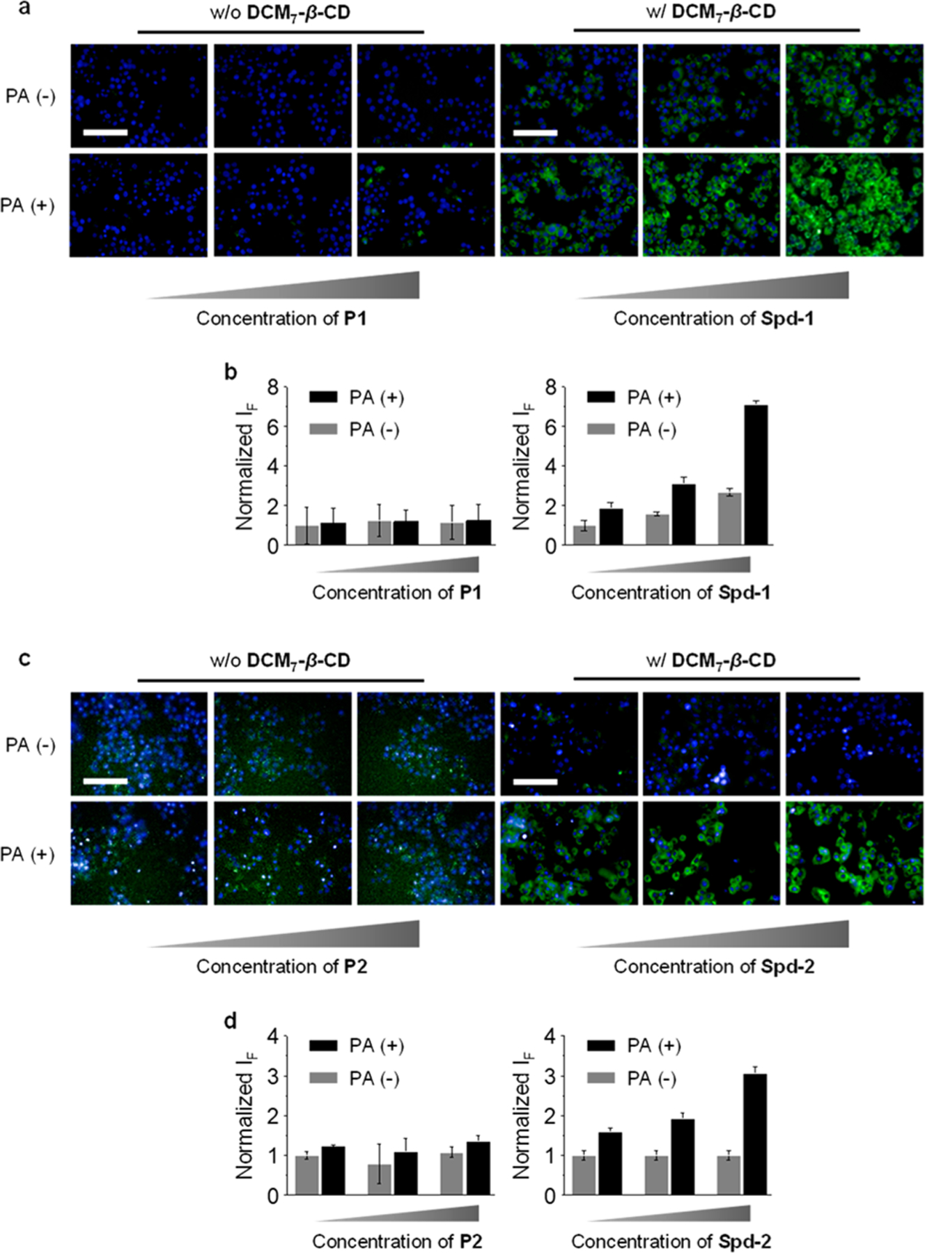
Dose-dependent fluorescence imaging (a) and quantification (b) of Hep-G2 cells (preincubated with 500 μM of palmitic acid (PA)) treated with P1 (0.2 μM, 0.5 μM, 1 μM) or Spd-1 (P1/DCM7-β-CD = 0.2/4 μM, 0.5/5 μM, 1/8 μM). Dose-dependent fluorescence imaging (c) and quantification (d) of Hep-G2 cells (preincubated with 500 μM of palmitic acid (PA)) treated with P2 (0.25 μM, 0.5 μM, 1 μM) or Spd-2 (P2/DCM7-β-CD = 0.25/4 μM, 0.5/5 μM, 1/8 μM). Excitation and emission channels for FITC are 460–490 and 500–550 nm, respectively. Cell nuclei were stained by Hoechst (excitation and emission channels are 360–400 and 410–480 nm, respectively). Scale bar = 100 μm (applicable to all images in panels and c).
