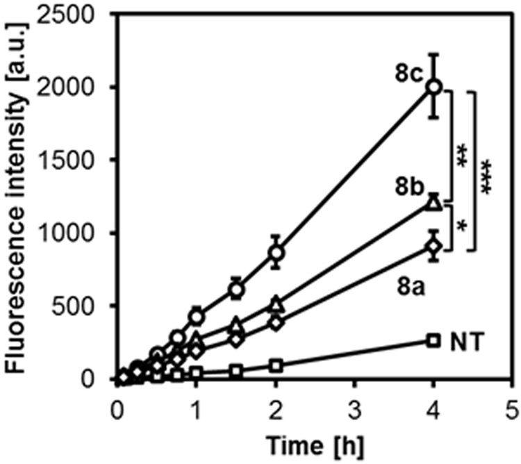
Figure 3.

H2S release in HUVECs. Cells in a 96 well plate were pretreated with the WSP-1 H2S detection probe in PBS for 30 min and cultured in medium without (NT, squares) or with micelles 8a (rhombus), 8b (triangles) and 8c (circles). ADT concentration: 50 μM. The fluorescence intensity was monitored using the bottom reading mode (λex=465 nm, λem=515 nm). *p < 0.05, **p < 0.01,***p < 0.001 (n=3).
