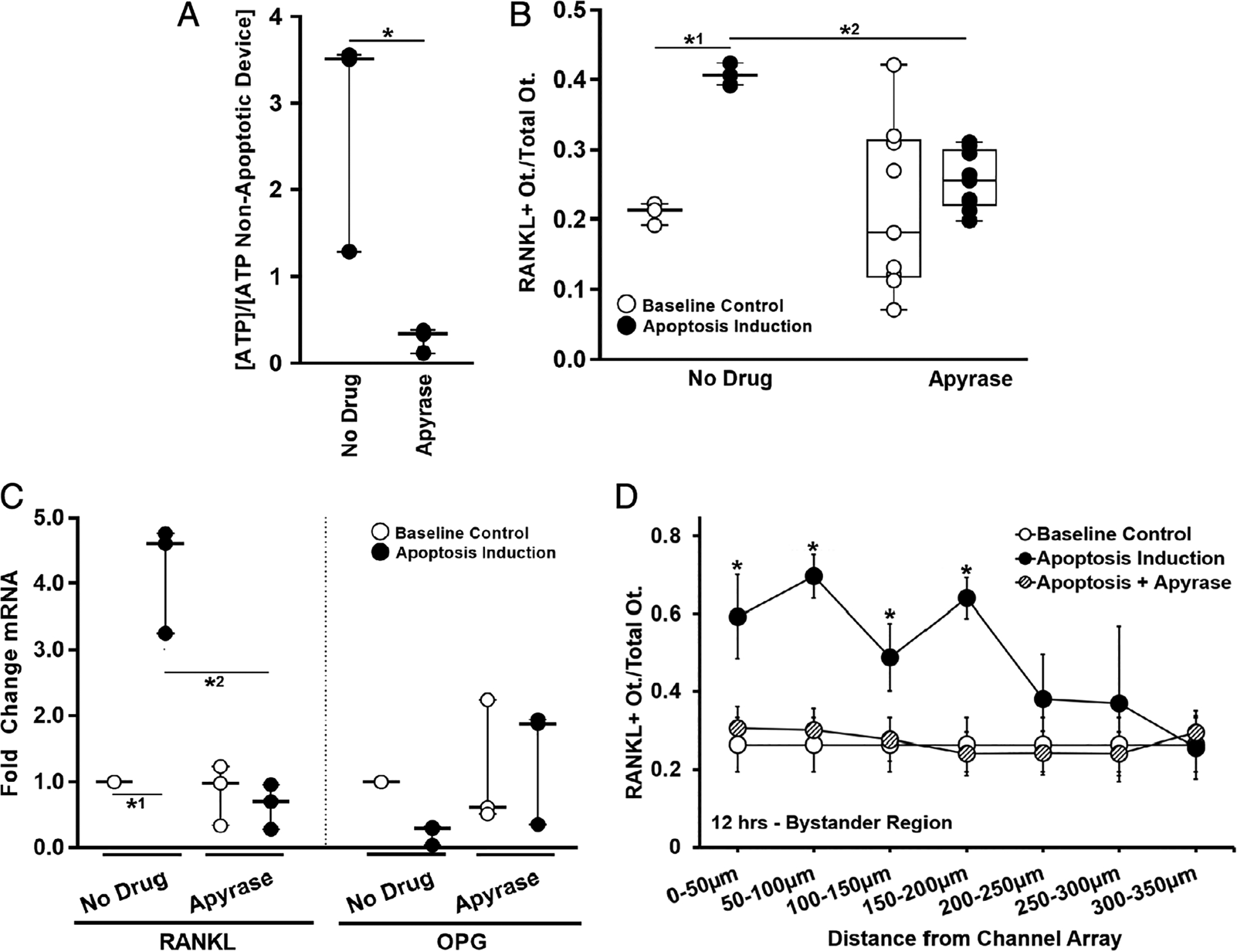
Fig. 4.

Effect of removing ATP on bystander osteocyte RANKL expression. (A) Induction of apoptosis induced sustained elevation in extracellular ATP in the Mμn (12 hours post-apoptosis induction, n = 3 devices, *p = .029); this increase was no longer seen when apyrase was added to the medium to hydrolyze the extracellular ATP. (B) The increased numbers of RANKL-expressing osteocytes expected after apoptosis induction (*1p = .031 versus control) was prevented by the addition of apyrase (p = .825 versus baseline control, *2p = .043 versus apoptotic control), n = 3 devices for control and n = 9 devices for apyrase treatment. (C) Scavenging the extracellular ATP with apyrase also prevented the changes in RANKL and OPG gene expression in bystander osteocytes caused by apoptosis of neighboring cells. (RANKL/apyrase p = .999 versus baseline control. *2p < .0001 versus no drug apoptotic control). n = 3 samples (three devices per sample), tested in triplicate. (D) Effect of apyrase on distribution of RANKL-expressing osteocytes as a function of the distance from the nanochannel array/apoptotic signal source, showing that hydrolyzing the ATP prevented increases in osteocyte RANKL expression through the entire bystander compartment (n = 9 devices, significance only for apoptosis induction-no apyrase versus baseline control [*p = .0001 at 0 to 50 μm, p = .0001 at 50 to 100 μm, p = .022 at 100 to 150 μm, and p < .0001 at 150 to 200 μm]). Data are shown as mean ± SD.
