Fig. 6.

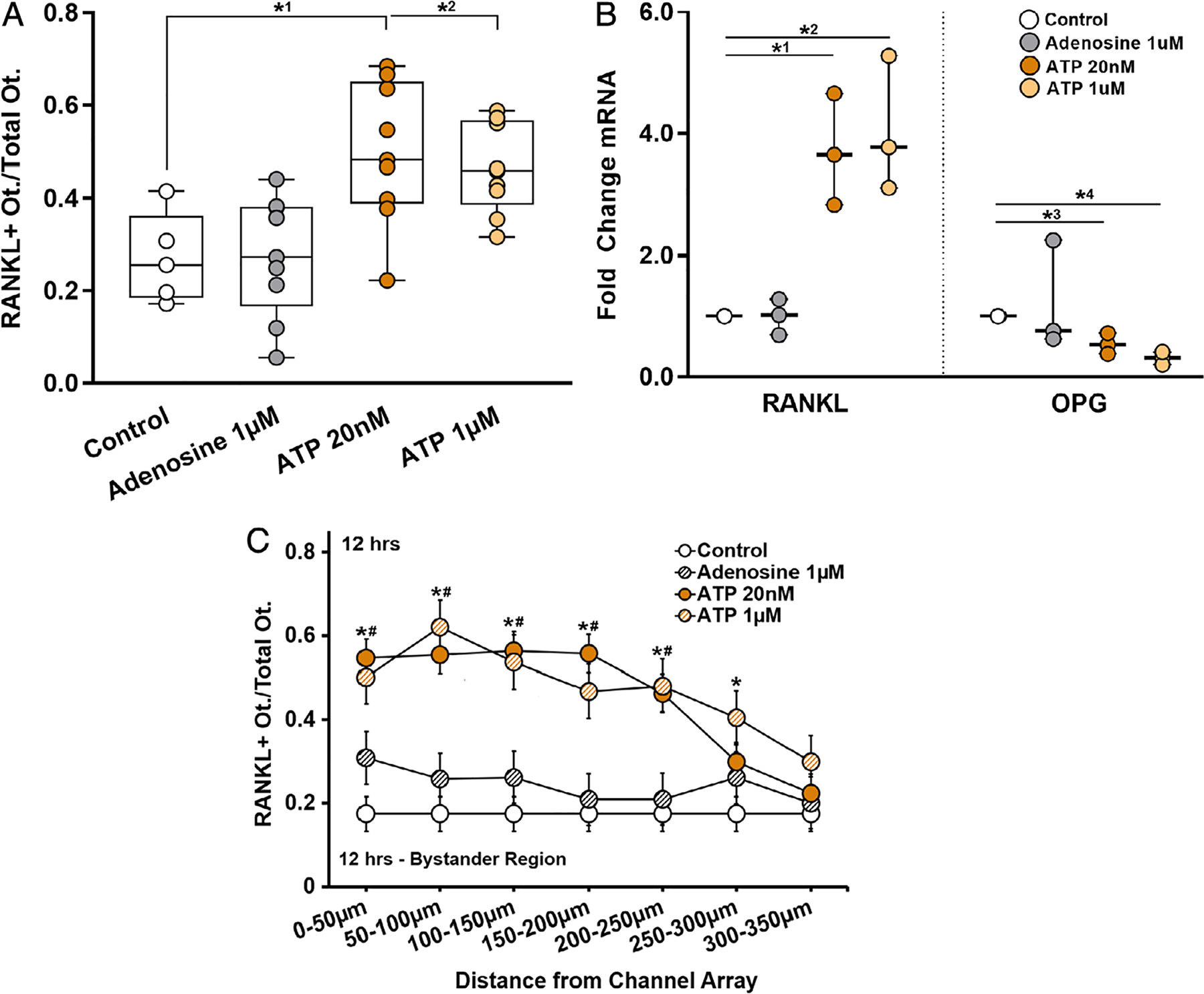
Purinergic stimulation induced changes on osteocyte RANKL expression. (A) ATP at 20nM and 1μM both similarly increased the number of RANKL-expressing osteocytes in the bystander compartment nearly twofold compared to control (*1p = .0133 and *2p = .044, respectively); adenosine treatment had no effect. n = 5 to 9 devices. (B) RANKL mRNA was markedly increased with exposure to ATP at 20nM or 1μM (*1p = .008 and *2p = .004) and OPG expression significantly decreased (*3p = .007 and *4p < .001, respectively), n = 3 samples (3 devices per sample), tested in triplicate; again adenosine had no effect. (C) The distribution of RANKL-expressing cells as a function of distance from the ATP source (nanochannel array) was similar to that observed when bystander osteocyte RANKL was triggered by apoptosis of neighboring cells; see Fig. 3. Both ATP 20nM* and 1μM# treatment groups were significantly different compared to untreated controls up to 300 μm from the ATP source (n = 9 devices), p = .001 at 0 to 50 μm, p = .0001 at 50 to 100 μm, p = .002 at 100 to 150 μm, p = .001 at 150 to 200 μm, p = .002 at 200 to 250 μm, and p = .011 at 250 to 300 μm). Adenosine had no effect. Data are shown as mean ± SD.
