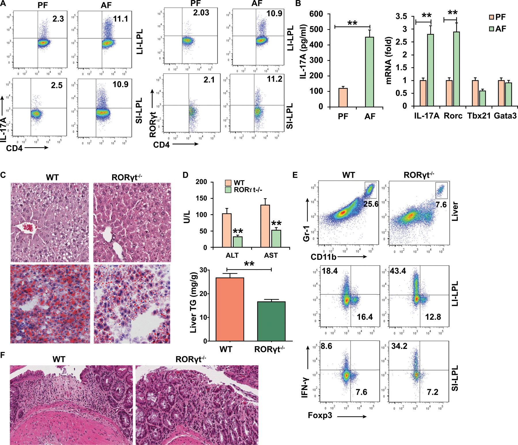
Figure 1. ALD is associated with a pro-inflammatory shift in intestinal immune cells.

WT mice (A-B) and RORγt−/− (C-F) mice were fed control (pair-fed, PF) or alcohol (alcohol-fed, AF) diet and sacrificed 15–22 days later.
(A) Intracellular staining of IL-17A and RORγt in CD4+ T cells in the lamina propia from large intestine (LI-LPL) or small intestine (SI-LPL).
(B) ELISA analysis of IL-17A in the supernatant of colonic LPLs stimulated with IL-23 (left) and real-time PCR analysis of IL-17A and Rorc mRNA in colonic LPLs (right).
(C-F) WT mice or RORγt−/− mice were fed alcohol diet and sacrificed 15 days later.
(C) HE and Oil red O staining of liver tissue.
(D) Serum levels of ALT and AST and hepatic triglycerides.
(E) Frequencies of immature myeloid cells (CD11b+Gr-1+) in the liver and Th1 and Tregs cells in the gut LPLs.
(F) Large intestine was stained by H&E.
Data in all panels are presented as Mean ± SEM. n=11 * P<0.05; ** P<0.01
