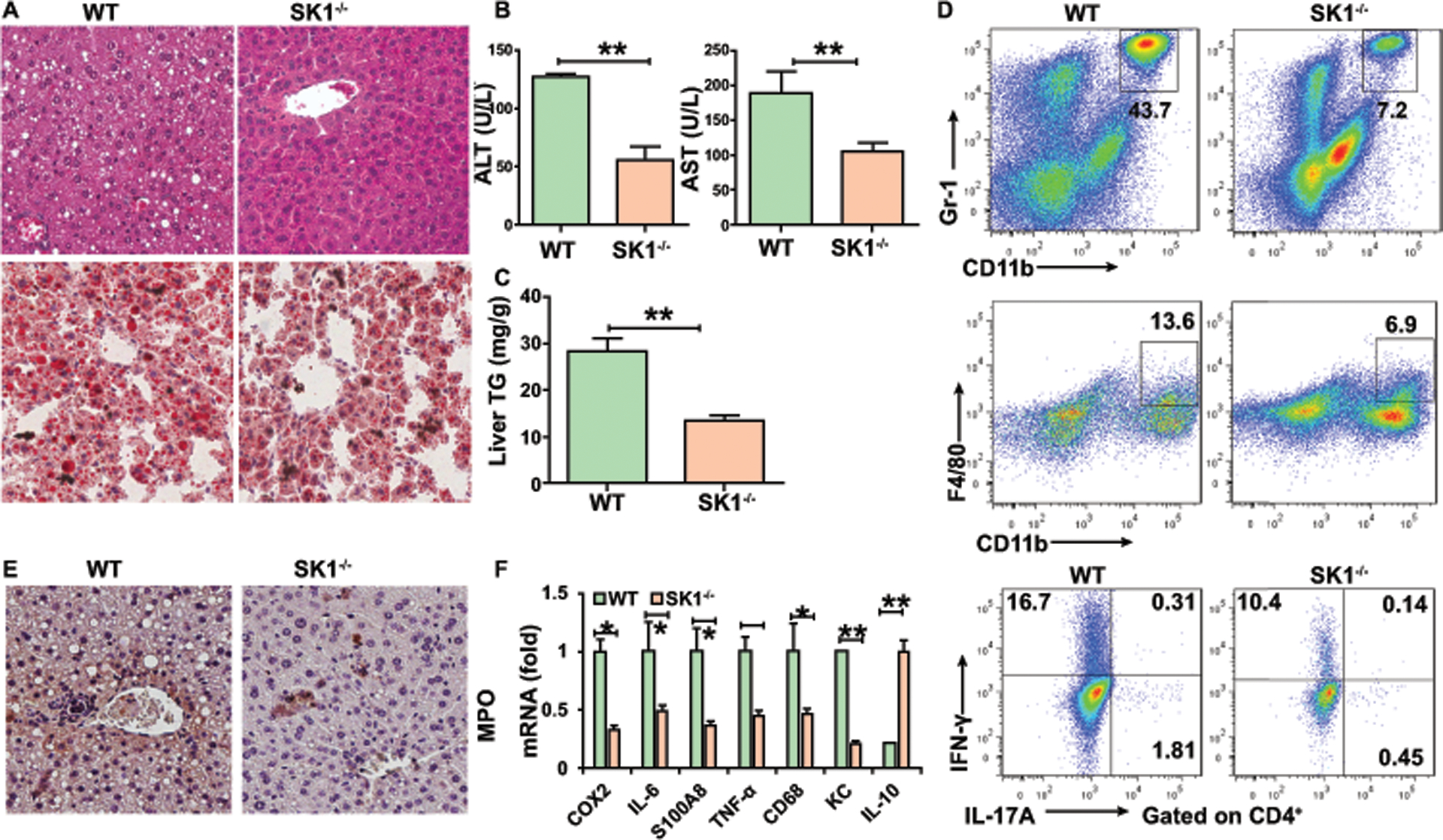
Figure 5. Deficiency of SK1 attenuates alcoholic liver inflammation, steatosis, and damage.

WT or SK1−/− mice were fed alcohol diet and sacrificed 15 days later.
(A) H&E staining and Oil red O staining of liver.
(B) Serum ALT and AST.
(C) Hepatic triglycerides.
(D) Frequencies of immature myeloid cells, macrophages, Th1, and Th17 cells in the liver.
(E) Immunohistochemistry staining of myeloperoxidase (MPO) in the liver.
(F) Real-time PCR analysis of the relative expression of indicated genes in the liver.
Data in all panels are presented as Mean ± SEM. n=11 * P<0.05; ** P<0.01
