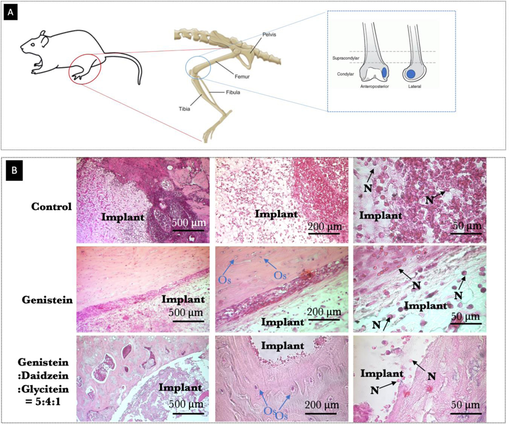
Fig. 10.

(A) Schematic representation of the surgical procedure and the 3D printed TCP scaffold (diameter of 3 mm and height of 5 mm) utilized for implantation (B) Optical microscopy images of decalcified tissue-implant specimens after H&E staining showing inflammatory cell recruitment after 24 days of surgery in rat distal femur model. Blue and black arrow show neutrophil recruitment and presence of osteocytes, respectively.
