Figure 3.
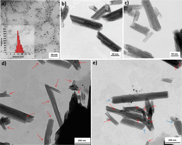
TEM micrographs of (a) as-synthesized SPION@OA (in the inset the SPION diameter distribution); (b–e) SPION-in-HNT prepared by prefunctionalization of HNT with TDP in the lumen. Panels (b) and (c) show images taken at higher magnifications compared to that used for (d) and (e). Red arrows mark the HNT containing SPION. Blue arrows indicate SPION at the edge of few HNT.

