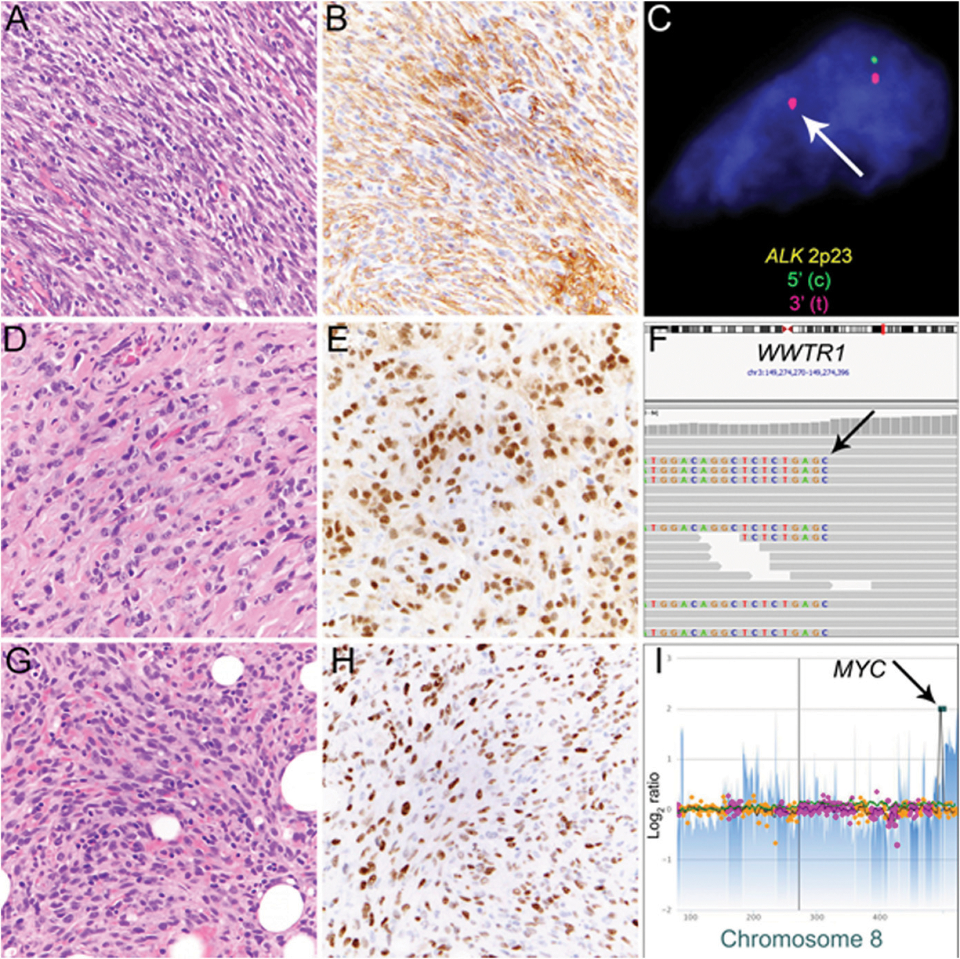
FIGURE 3. Mesenchymal Neoplasms With Distinct Cytogenetic Aberrations and Associated Immunohistochemical Markers.

Inflammatory myofibroblastic tumor is characterized by a population of spindled tumor cells arranged in fascicles with scattered inflammatory cells (A) and expression of ALK in tumor cells (B). Rearrangement of the ALK locus at 2p23 can be detected by fluorescence in situ hybridization (C; break-apart signal indicated by arrow). Malignant epithelioid hemangioendothelioma (EHE) consisting of strands of tumor cells with epithelioid morphology, nuclear atypia, and glassy amphophilic cytoplasm embedded in a myxohyaline to collagenous stroma (D). Most cases of EHE have diffuse nuclear expression of CAMTA1 (E) resulting from WWTR1-CAMTA1 fusion. In this case, next-generation sequencing detected rearrangement involving the WWTR1 coding as evidenced by split reads (F, arrow) that match to CAMTA1 (not shown). Postradiation angiosarcoma of the breast consists of atypical endothelial cells growing in strands and sheets and diffusely infiltrating preexisting fat (G) is characterized by nuclear expression of MYC (H) resulting from high-level MYC amplification (I, arrow).
