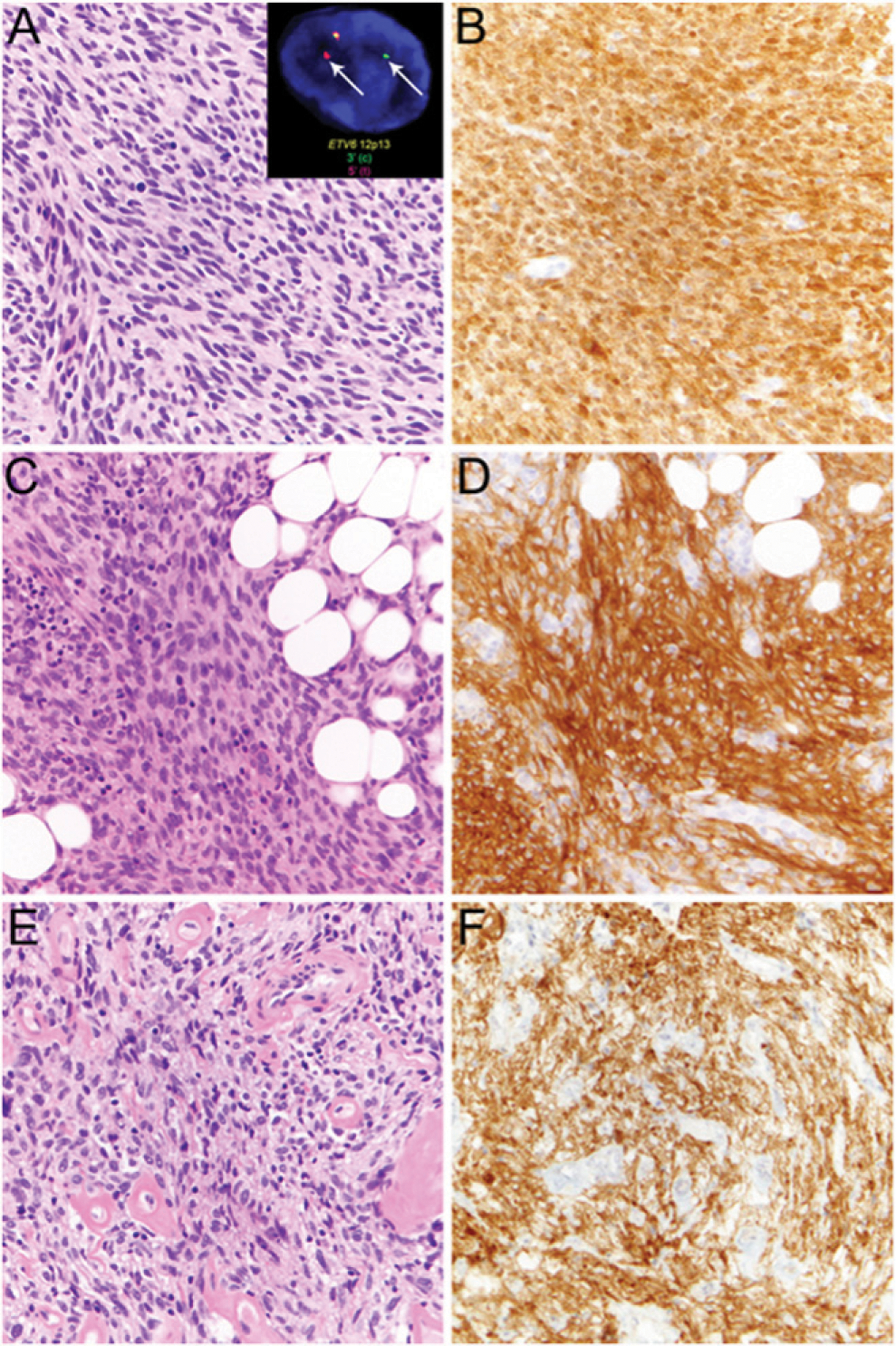
FIGURE 4. Examples of Mesenchymal Neoplasms Showing Pan-TRK Expression by Immunohistochemistry.

Infantile fibrosarcoma comprises monotonous population of spindle cells (A) with ETV6-NTRK3 rearrangement, detected by ETV6 break-apart fluorescence in situ hybridization (A, inset, arrows), and shows diffuse staining by pan-TRK immunohistochemistry (B). Lipofibromatosis-like neural tumor with spindled tumor cells lacking nuclear atypia containing wavy neural-like nuclei and eosinophilic cytoplasm with diffuse infiltration of adjacent fat (C) shows diffuse pan-TRK staining (D). Unclassified sarcoma of the endocervix comprising ovoid to spindled tumor cells with nuclear atypia scattered in a collagenous stroma with prominent hyalinized blood vessels (E) showing positive pan-TRK staining (F) suggestive of underlying NTRK rearrangement.
