
FIGURE 5. MRI-Guided Radiotherapy for Sarcoma Liver Metastasis.
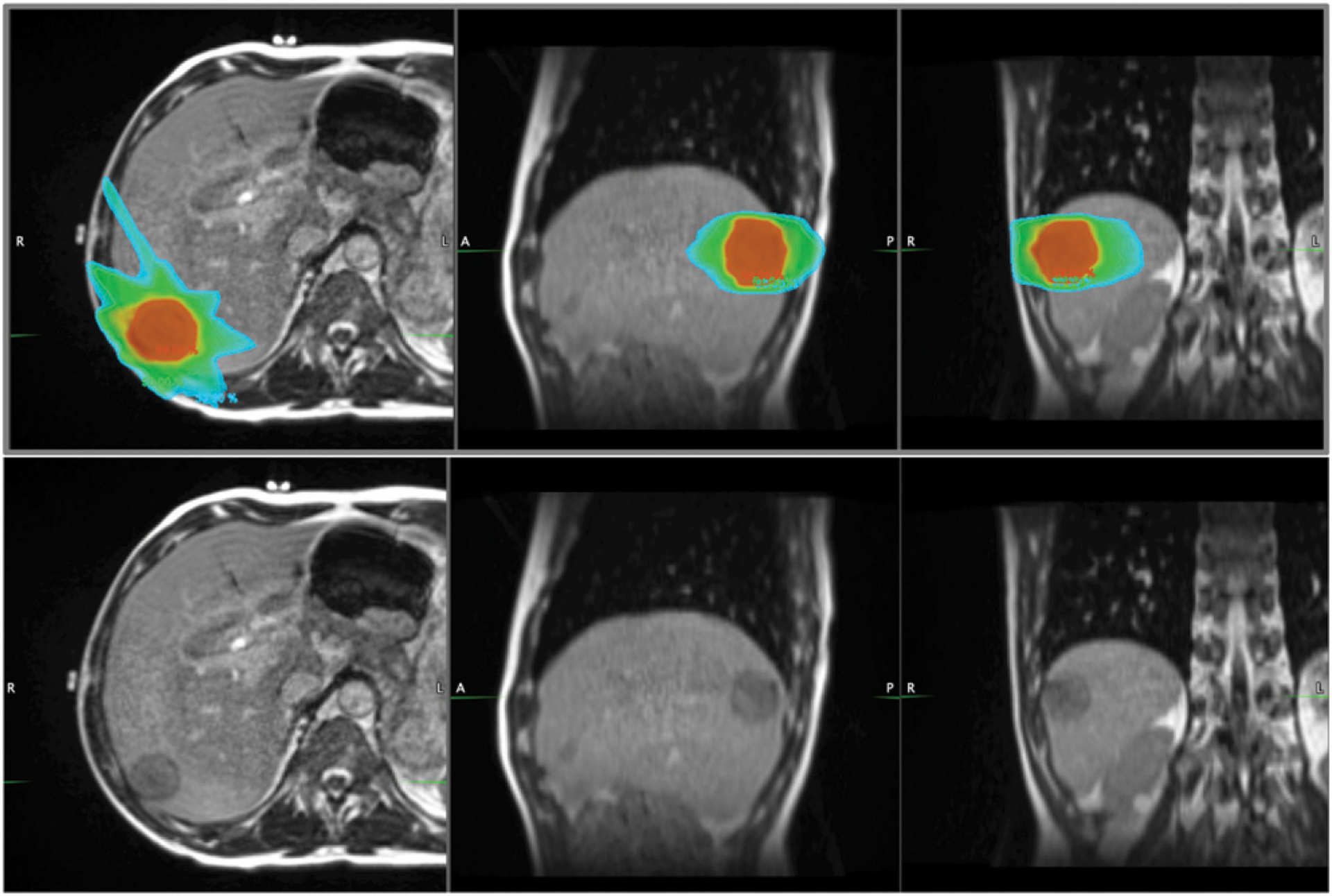
Radiation dose distribution (top panel) for a patient receiving magnetic resonance (MR)-guided stereotactic body radiotherapy for oligometastatic spindle cell sarcoma of the liver. A 172s balance steady-state free precession MR sequence was used to acquire three-dimensional anatomic MRI with 1.5 mm isotropic resolution using MRIdian’s 0.35T on-board MRI. There are distinct advantages of MR-guided radiotherapy for sarcoma at simulation, treatment planning, and patient setup prior to treatment delivery. In this case, the target lesion was not identifiable on CT imaging. The ability to visualize the tumor just prior to treatment allowed safe reduction of the planning margin from 1 cm to 5 mm, reducing radiation dose to surrounding normal tissue. Given the challenging treatment setup for sarcoma extremity lesions, the added imaging allowed more reproducible patient positioning. From left to right in both top and bottom panels: axial, sagittal, and coronal MR images. Isodose colors (top panel): red, 99%; green, 50%, light blue, 33%.