
Figure 3. Deletion of the MYH7-C3 enhancer increases MYH6 and reduces MYH7 mRNA and protein and produces faster contraction and relaxation in engineered heart tissues.
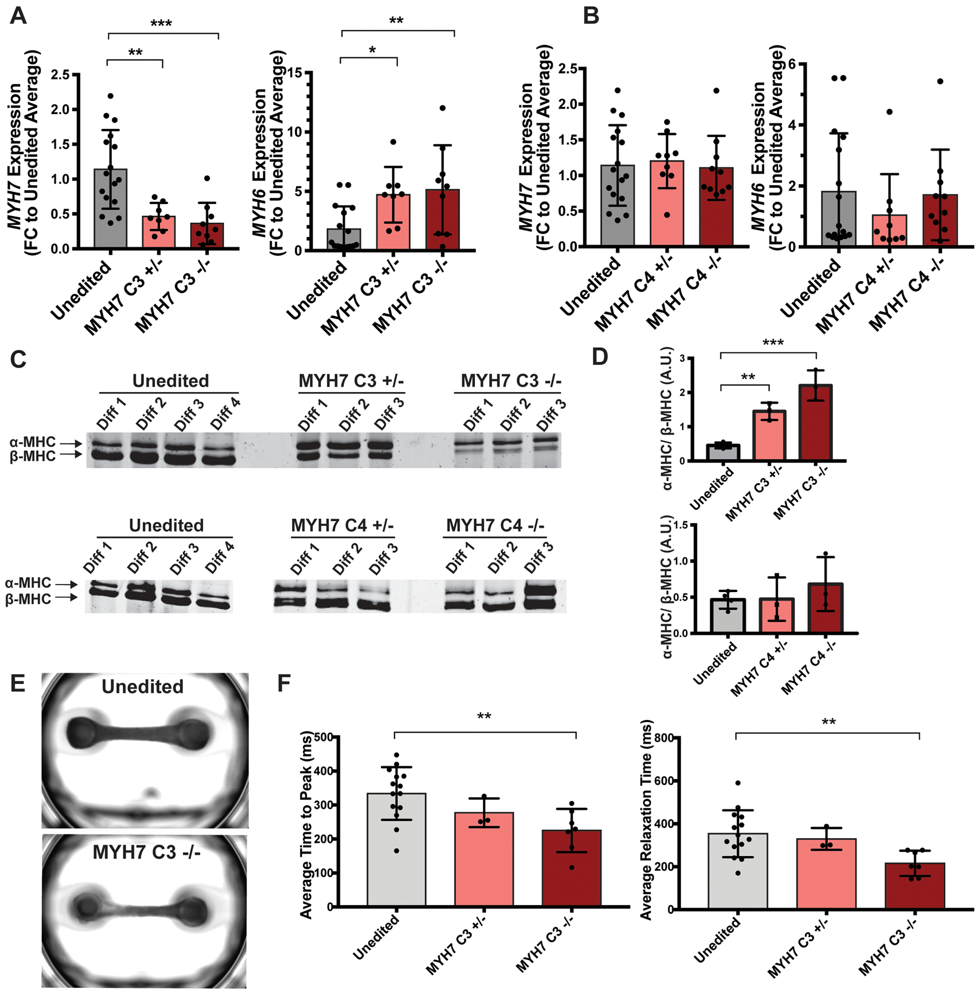
A. Gene editing was used to delete the MYH7-C3 enhancer heterozygously (+/−) or homozygously (−/−). MYH6 and MYH7 mRNA expression was assayed by qPCR and showed a dose-dependent increase in MYH6 expression and reduction in MYH7 expression. Therefore, the MYH7-C3 enhancer is required for MYH7 expression (unedited n=16, MYH7 C3+/− n=8, MYH7 C3−/− n=9.) B. Deletion of the MYH7-C4 enhancer had little effect, demonstrating a specificity of these findings to MYH7-C3 (unedited n=16, MYH7 C4+/− n=9, MYH7 C4−/− n=10.) C. α-MHC and β-MHC protein ratios were quantified using SDS-PAGE. D. Quantification of α-MHC/β-MHC protein ratios demonstrating correlation with the differences seen at the RNA level. E. Representative images of engineered heart tissues (EHTs) containing unedited or MYH7-C3 homozygous deleted IPSC-CMs. F. Average time to peak (top) and average relaxation times (bottom) measurements of EHT contractions containing unedited or MYH7-C3 deleted cells showed a decrease in time to peak contraction and relaxation in MYH7-C3 deleted EHTs, consistent with the shift from MYH7/β-MHC to MYH6/α-MHC and the known faster ATPase cycle for α-MHC. Each point represents the average time to peak measurement of a single EHT across multiple contractions (unedited n=14, MYH7 C3+/− n=3, MYH7 C3−/− n=7.) All data shown as mean ±SD. * determined by one-way ANOVA with Dunnett’s multiple comparisons correction. *<0.03, **<0.0021, ***<0.0002.