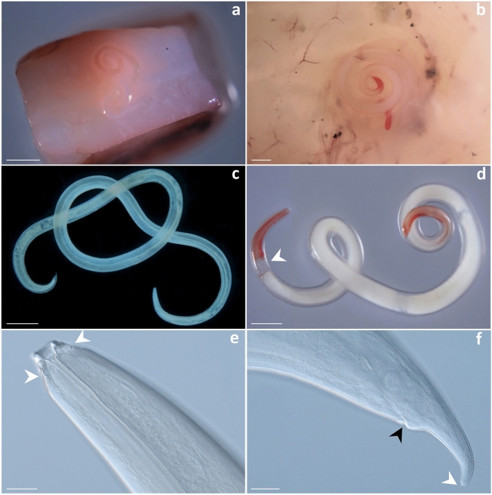
Figure 1.
Lappetascaris sp. and Anisakis physeteris in Histioteuthis squids collected from the Tyrrhenian Sea. Lappetascaris larva in the mantle musculature of an umbrella squid (a). Anisakis physeteris in the testis of an umbrella squid. Note the worm extremities reddish in colour (b). Microscopic view of Lappetascaris sp. larva (c) and A. physeteris showing the extremities reddish in colour and ventriculus (arrow) (d). Cephalic extremity of Lappetascaris sp. larva showing sclerotized formations (arrows) (e). Caudal extremity of Lappetascaris sp. larva showing anus (black arrow) and cuticular spike (white arrow) (f). Scale bar: 2000 µm (a); 1000 µm (b,c,d); 50 µm (e,f).

