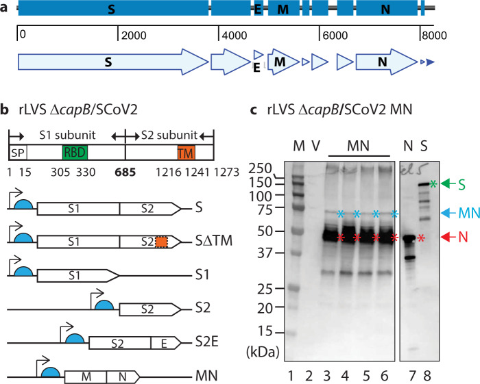
Fig. 1. Construction of rLVS ΔcapB/SARS-CoV-2 vaccines.
a Schematic of SARS-CoV-2 genomic region encoding four major structural proteins, Spike (S) glycoprotein, Envelope (E), Membrane (M), and Nucelocapsid (N) protein. b Diagram of S protein and the antigen expression cassettes for S, SΔTM, S1, S2, fusion protein of S2 and E (S2E) and fusion protein of M and N (MN) downstream of the F. tularensis bacterioferritin (FTT1441) promoter (Pbfr) (thin black arrow) and Shine-Dalgarno sequence (light blue half circle). SP, signal peptide for S protein; RBD, receptor-binding domain; and TM, transmembrane domain. c Protein expression of rLVS ΔcapB/SCoV2 MN. Total bacterial lysates of 4 clones of rLVS ΔcapB/SCoV2 MN (lanes 3–6, as indicated at the bottom of the left panel) were analyzed by SDS-PAGE and Western blotting with an anti-SARS-CoV-1 guinea pig polyclonal antibody (BEI Resources, NR-10361), which readily detected the full-length MN (~75 kDa, less abundant), indicated by blue asterisks to the right of the protein bands, and the highly abundant breakdown product N protein (~46 kDa), indicated by red asterisks to the right of the protein bands. The anti-SARS-CoV-1 guinea pig polyclonal antibody also detected the N (red arrow and asterisk) and S (green arrow and asterisk) proteins of SARS-CoV-1 (lanes 7 and 8), which served as positive controls. V, LVS ΔcapB vector (lane 2). The left and right panels are from the same gel (Supplementary Fig. 6). The sizes of the molecular weight markers (M) are labeled to the left of the panels.

