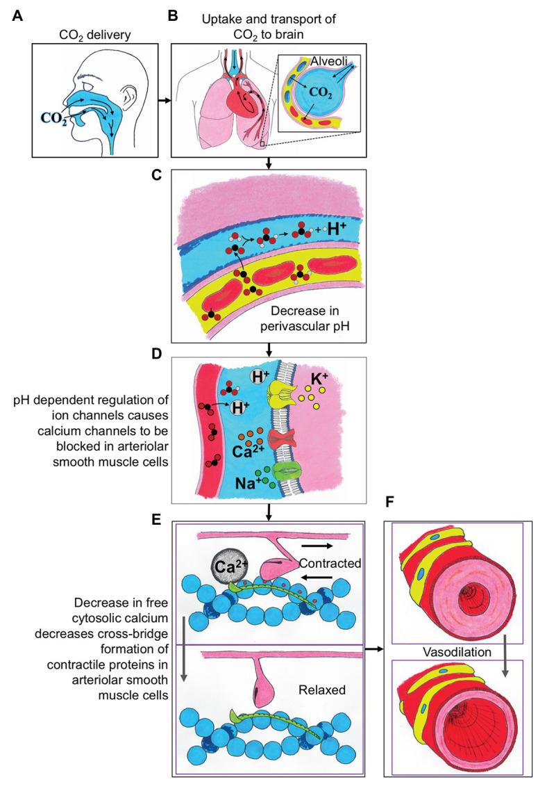
Figure 1.

A schematic describing the integrative physiologic mechanisms governing cerebrovascular reactivity (CVR) in response to elevated carbon dioxide (CO2) stimuli. (A) The CO2 is delivered to the participant via mouth or nose. (B) The uptake of CO2 is in the alveoli of the lungs, where the gas is exchanged into the blood, and then transported to the brain. (C) The CO2 exchanges from the blood into the perivascular space and causes a decrease in perivascular pH. (D) The decreased perivascular pH causes the calcium channels to be blocked in arteriolar smooth muscle cells. (E) Decreased local calcium concentration results in relaxation of arteriolar smooth muscle cells. (F) The relaxation of arteriolar smooth muscle cells leads to local vasodilation and subsequent CVR contrast. H+, proton; Ca2+, calcium; Na+, sodium; and K+, potassium.
