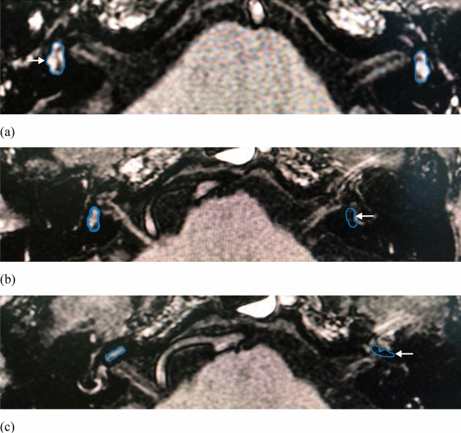
Figure 1.
Three-dimensional fluid attenuated inversion recovery (3D-FLAIR) images of patients who diagnosed with definite MD. (a) The region of interest (ROI) placement for the mild endolymphatic hydrops (grade 1) in right-sided vestibule (white arrow). (b) The ROI placement for the obvious endolymphatic hydrops (grade 2) in left-sided vestibule (white arrow). (c) The ROI placement for the significant endolymphatic hydrops (grade 2) in left-sided cochlea (white arrow).

