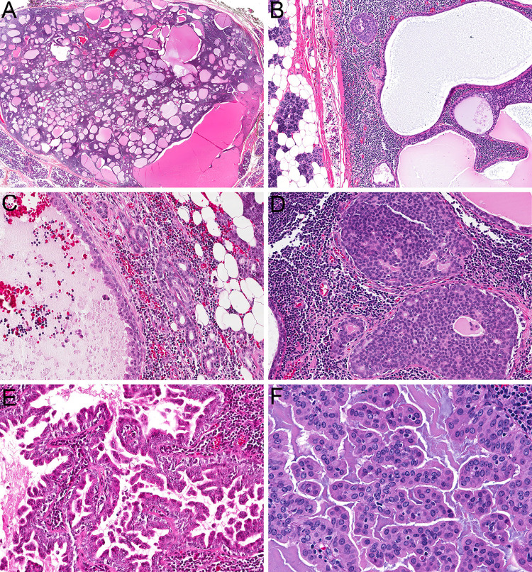
Fig. 1.
The tumors were well-circumscribed and consisted of a complex epithelial proliferation embedded in dense lymphoid stroma (a, 2x). A well-developed lymph node capsule with sinus histiocytes was recognizable at the periphery of all lesions (b, 10x). Benign salivary inclusions were also present within the lymph node parenchyma adjacent to the tumor cells in three cases (c, 10x). Tumors showed a variety of architectural patterns, including cribriform (d, 20x), cystic, and micropapillary growth (e, 20x). Tumor cells were bland with a moderate amount of eosinophilic cytoplasm and oval nuclei with delicate chromatin and inconspicuous nucleoli (f, 40x)

