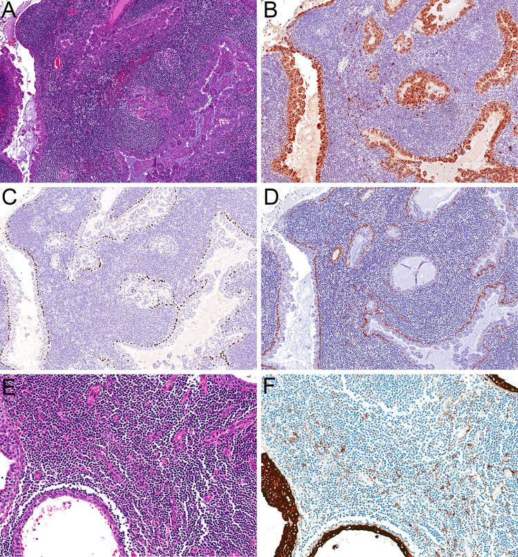
Fig. 2.
Immunohistochemistry confirmed classification as the intercalated duct subtype of IDC (a, 20x), with tumor cells diffusely positive for S100 protein (b, 20x) and an intact myoepithelial layer, positive with p40 (c, 20x) and SMA (d, 20x). Immunohistochemistry demonstrated that the lymphoid stroma represented lymph node parenchyma rather than tumor-associated lymphoid proliferation (e, 20x) with positivity for low molecular-weight cytokeratin Cam 5.2 in extrafollicular reticulum cells (f, 20x)

