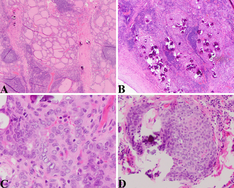
Fig. 2.
DSV-PTC: a Low magnification view of this tumor showing an area with a heavy lymphocytic infiltrate, thick bands of fibrous tissue that divide the thyroid into lobules, and numerous psammoma bodies. This is a deceptive field of view given that the thyroid tissue that is visible is non-neoplastic thyroid (HE 40X). b Numerous psammoma bodies characteristic of this aggressive variant of PTC. Here in this field the psammoma bodies are concentrated in the more solid appearing and neoplastic foci (HE 40X). c High magnification view showing the characteristic nuclear morphology of PTC; nuclear crowding, pleomorphism, central clearing, and nuclear grooves (HE, 400X). d Solid squamous area with psammoma bodies (HE, 200X)

