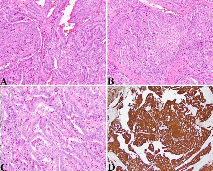
Fig. 3.
CMV-DTC: a Medium magnification view of CMV-PTC showing a cribriform pattern in the left half of the image, a more papillary pattern in the right half and a small central morule (HE, 100X). b Medium magnification view showing a solid squamous morula embedded in a cribriform pattern of PTC (HE, 100X). c Higher magnification showing an area with typical nuclear features of PTC (crowding, elongated and enlarged nuclei, grooved nuclei, nuclear clearing) (HE, 200X). d Beta-catenin stain shows diffuse nuclear and cytoplasmic staining (Beta-catenin, 40X)

