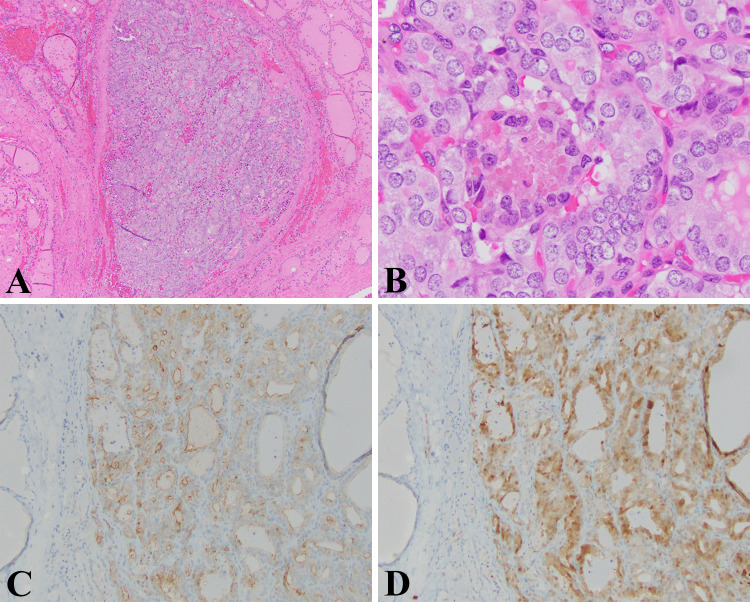
Fig. 5.
NIFTP: a Low magnification view of a minimally encapsulated but well-circumscribed follicular lesion surrounded by normal thyroid parenchyma (HE, 40X). b The nuclei clearly demonstrated morphological features of papillary thyroid carcinoma. In this field, we see mild nuclear anisocytosis, focal chromatin clearing, crowding with overlapping, nuclear grooves (HE, 400X). c The tumor cells mostly express HBME-1 in the characteristic membranous pattern (HBME-1, 100X). d Similarly, the tumor shows nearly diffuse expression of Galectin-3 with appropriate cytoplasmic and nuclear staining (Galectin-3, 100X)

