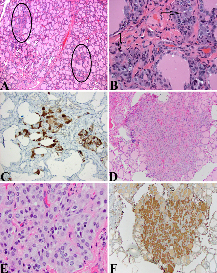
Fig. 6.
CCH and MTC: a Thyroidectomy specimen from the 3-year-old boy presented above showing vague hypercellular areas having a grayish tinge intercalated within the thyroid parenchyma (ovals) (HE, 40X). b High magnification view of one of the highlighted areas in (A) showing large cells with relatively indistinct nuclei and abundant amphophilic cytoplasm in clusters and around follicles. While the nuclei are relatively indistinct even compared to normal thyroid follicular epithelial cells, they are quite noticeable given that under normal circumstances, C-cells are difficult to ascertain on routine HE sections. These features qualify for cytological atypia and should be considered as such (HE, 400X). c These cells noted in (b) are easily highlighted by a calcitonin stain (Calcitonin, 200X). d Small relatively uncircumscribed lesion in an adolescent with MEN2A. This cellular lesion could be mistaken for an intrathyroidal parathyroid gland or small hyperplastic thyroid epithelial cell-derived nodule (HE, 40X). e The cells have a “neuroendocrine” appearance and form small organoid nests. The cytological features include small uniform nuclei, bland chromatin, and amphophilic cytoplasm (HE, 400X). f A calcitonin stain again confirms the ontogeny from C-cells and this lesion as a medullary thyroid microcarcinoma in a patient undergoing prophylactic thyroidectomy for MEN2A (Calcitonin, 40X)

