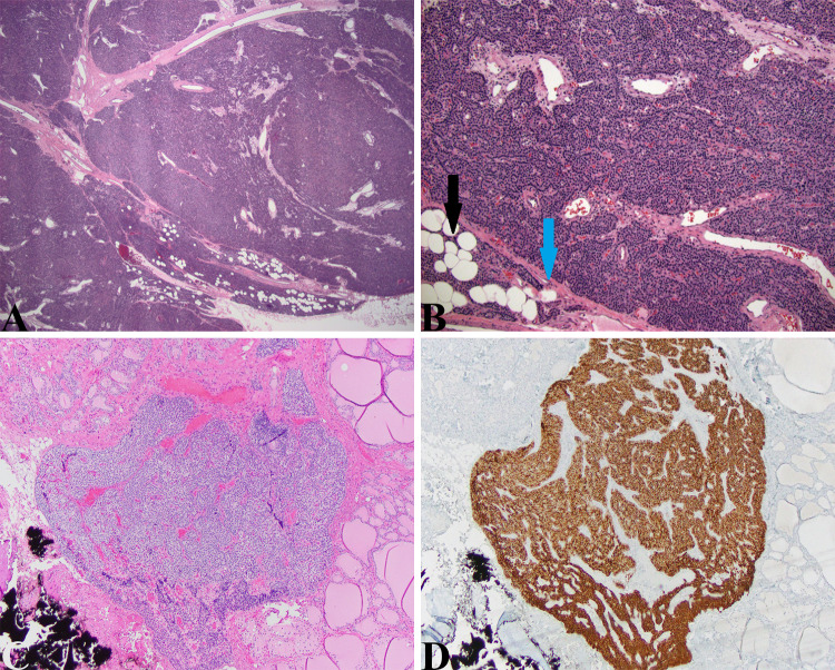
Fig. 8.
Parathyroid adenoma and parathyroid gland: a Large parathyroid adenoma
taken from the 11-year-old boy presented above. This uniformly hypercellular gland is finely encapsulated (blue arrow in (b)) and abuts normal parathyroid tissue characteristic of adenomas (black arrow in (b)). Some thicker fibrous bands are seen traversing the adenoma proper but other features to suggest malignancy (capsular/vascular invasion, trabecular growth, atypia, atypical mitoses) were not seen (HE, 20X, 40X). c Intrathyroidal lesion seen in the thyroidectomy specimen from the young girl with MEN2A presented above that has similar cytological features to that of the medullary thyroid microcarcinoma. This lesion is clearly parathyroid gland confirmed by the diffuse staining with anti-PTH antibody in (d) (HE, 40X and PTH, 40X)

