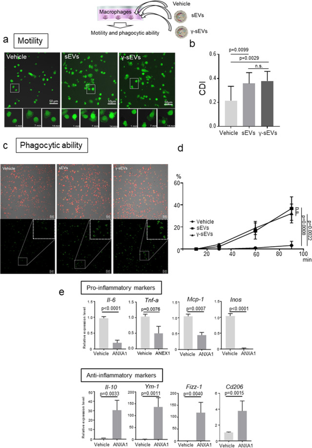
Fig. 2. Macrophage motility and phagocytic ability after addition of AD-MSC-sEVs (sEVs) or AD-MSC-γ-sEVs (γ-sEVs).
a Motility of GFP+ macrophages after addition of vehicle, sEVs, and γ-sEVs (Supplementary Movie 1; vehicle, sEVs and γ-sEVs from left to right). b Quantification of morphological changes in macrophages. Data are presented as means ± SD; ten screens were counted in each experiment. CDI, cell deformation index. c Phagocytic ability of DsRed+ macrophages after phagocytosis of pHrodo™ Green Zymosan Bioparticles™ conjugate (Supplementary Movie 2; vehicle, sEVs, and γ-sEVs from left to right). d Frequencies of green fluorescent macrophages observed using time-lapse imaging. Data are presented as means ± SD; ten screens were counted in each experiment 90 min after the addition of sEVs or γ-sEVs. Scale bar = 50 μm. e Change in macrophage polarity after treatment with recombinant human annexin-A1 (ANXA1).

