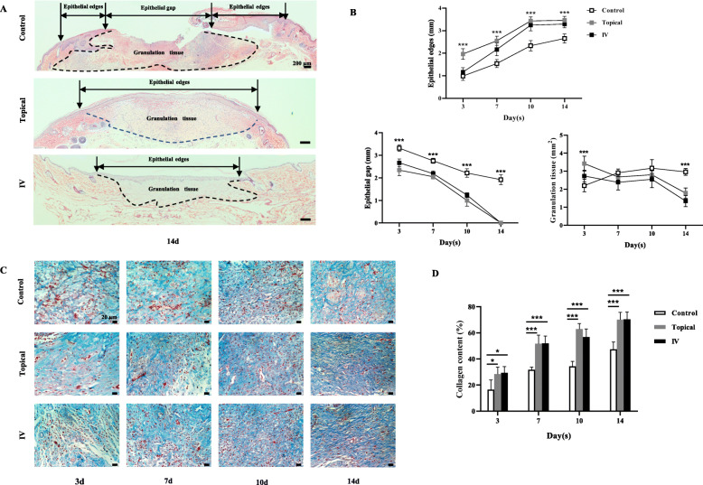
Fig. 4.
Epithelialization and granulation tissue regeneration assessment in rats treated topically and systemically. a H&E-stained sections of wound specimens from rats at days 14 (4 × 10). Note that the picture showed the analytical method of the length of epithelial edges, the length of the epithelial gap, and the area of granulation tissue. b The length of epithelial edges, the length of the epithelial gap, and the area of granulation tissue at days 3, 7, 10, and 14. c Collagen deposition was assessed by Masson’s trichrome (40 × 10) at days 3, 7, 10, and 14. d The collagen content at days 3, 7, 10, and 14. Values are mean ± SD. *P < 0.05, **P < 0.01, ***P < 0.001

