Abstract
Objective:
To evaluate the value of using low energy (keV) images in renal dual-energy spectral CT angiography (CTA) and adaptive statistical iterative reconstruction (ASIR) to reduce contrast medium dose.
Methods:
40 patients with renal CTA on a Discovery CT750HD were randomly divided into two groups: 20 cases (Group A) with 600 mgI kg−1 and 20 cases (Group B) with 300 mgI kg−1. The scan protocol for both groups was: dual-energy mode with mA selection for noise index of 10 HU, pitch 1.375:1, rotating speed 0.6 s/r. Images were reconstructed at 0.625 mm thickness with 40%ASIR, Group A used the conventional 70keV monochromatic images, and Group B used monochromatic images from 40 to 70 keV at 5 keV interval for analysis. The CT values and standard deviation (SD) values of the renal artery and erector spine in the plain and arterial phases were measured with the erector spine SD value representing image noise. The enhancement degree of the renal artery (ΔCT = CT(arterial) -CT(plain)), signal-to-noise ratio (SNR=CTrenal-artery/SDrenal-artery) and contrast-to-noise ratio (CNR=(CTrenal-artery-CTerector spine)/SDerector-spine) were calculated. The single factor analysis of variance was used to analyze the difference of ΔCT, SNR and CNR among image groups with p < 0.05 being statistically significant. The subjective image scores of the groups were assessed blindly by two experienced physicians using a 5-point system and the score consistency was compared by the κ test.
Results:
Contrast medium dose in the 300 mgI kg−1 group was reduced by 50% compared with the 600 mgI kg−1 group, while radiation dose was similar between the two groups. The subjective scores were 4.00 ± 0.65, 4.50 ± 0.60 and 3.70 ± 0.80 for images at 70 keV (600 mgI kg−1 group), 40 keV (300 mgI kg−1 group) and 45 keV (300 mgI kg−1 group), respectively with good consistency between the two reviewers (p > 0.05). The 40 keV images in the 300 mgI kg−1 group had similar ΔCT (469.77 ± 86.95 HU vs 398.54 ± 73.68 HU) and CNR (15.52 ± 3.32 vs 18.78 ± 6.71) values as the 70 keV images in the 600 mgI kg−1) group but higher SNR values (30.19 ± 4.41 vs 16.91 ± 11.12, p < 0,05)
Conclusion:
Contrast dose may be reduced by 50% while maintaining image quality by using lower energy images combined with ASIR in renal dual-energy CTA.
Advances in knowledge:
Combined with ASIR and energy spectrum, can reduce the amount of contrast dose in renal CTA.
Introduction
Computed tomography angiography (CTA) is commonly used for the accurate and non-invasive evaluation of the renal vascular anatomy. and the diagnosis of renal vascular lesions.1 Typical indications include the evaluation of suspicious renovascular hypertension, vasculitis, tumors, vascular malformations, and kidney structural diseases. The image quality of renal CTA depends on the iodine concentration in the renal artery lumen.2 The higher the iodine content, the better image comparison between the renal artery and the surrounding tissues.3
However, improving the image quality of CTA by increasing the dose of contrast agent will lead to a series of clinical problems, such as contrast-induced nephropathy (CIN).4 CIN is an acute kidney injury that occurs after the administration of contrast media, and is usually defined as an increase in serum creatinine (Scr) level ≥0.5 mg dl−1 or a 25% increase from the baseline value, assessed at 48 h after contrast exposure.5 The incidence of CIN ranges from 0 to 21%, depending on patient comorbidities, procedure type, route of iodine contrast medium administration [intraarterial (IA) vs intravenous (i.v.)], type of contrast agent, and so on.6 Several risk factors, including older age (>75 years), use of nephrotoxic medications, hypovolemia, higher contrast volume, chronic kidney disease, and intraarterial administration are associated with an increased risk of CIN.7,8 A recent meta-analysis showed that there is no exact relationship between the occurrence of CIN and the type of contrast agent and the route of contrast agent injection.9 The details of total contrast volume, operation time and contrast injection rate are often overlooked in CIN-related studies.
Therefore, developing the means to reduce patient contrast dose has become an urgent issue for clinicians and radiologists. Contrast dose reduction may cause reduced enhancement degree in vessels, and one of the effective ways to compensate for the decreased enhancement is to apply low tube voltages in CT scanning or low photon energies in image reconstruction. However, the use of low tube voltages or low photon energies may reduce the detection efficiency and increase noise in the images. Iterative reconstruction (IR) has been used in the “double-low dose (low radiation dose and low contrast agent dose)” CT to reduce image noise and to meet the diagnostic requirements, and it has been employed in CT diagnosis of the chest, abdomen, blood vessels, and other areas.10–12 As an iterative reconstruction applied in the projection data space based on noise models,13 the adaptive statistical iterative reconstruction (ASIR) is widely used clinically. Recent research data have demonstrated that ASIR has a potential in reducing the radiation dose, improving the image quality, and lesion detection.14–16 On the premise of obtaining satisfactory image quality, ASIR can reduce the radiation dose by 25–40% compared with the filtered back-projection (FBP) reconstruction.16,17 However, it is still unclear about the effectiveness of ASIR in a low-contrast CTA to evaluate kidney vasculatures when combined with low photon energy images.
Therefore, the purpose of this study was to explore the clinical application of ASIR algorithm with combination of low energy (keV) images in renal dual-energy CTA for patients with smaller than normal body-mass-index (BMI ≤24 kg/m2) that used low contrast agent concentration for reducing contrast dose.
Methods
Patients
In this prospective study, all research procedures were approved by the Ethics Committee of the Affiliated Hospital, Shaanxi University of Traditional Chinese Medicine, China. A written informed consent was obtained from all patients or their legal representatives for inclusion in the study. A total of 40 patients suspected of a renovascular disease were subjected for renal CTA in our hospital (Affiliated Hospital of Shaanxi University of Chinese Medicine) from May 2019 to December 2019, including 26 males and 14 females.
The inclusion criteria were as follows: patients needing renal artery examination, but without known renal artery disease; patients with body mass index (BMI) ranges from 18.5 to 23.9 kg m−2 and ages between 18 and 80 years. The exclusion criteria were as follows: patients at risk of being allergic to iodine contrast agents; patients with known renal insufficiency or severe heart failure or liver insufficiency.
40 patients were finally enrolled in the study and were randomly divided into two groups (Group A and B), with 20 cases in each group. There were 15 males and 5 females in Group A, with an average age of 56.75 ± 9.87 years and BMI of 20.61 ± 2.95 kg m−2. There were 16 males and 4 females in Group B, with an average age of 61.35 ± 10.08 years and BMI of 20.74 ± 3.56 kg m−2. All patients were fully conscious and were able to comply thoroughly with the commend during the CT examination. These patients exhibited normal levels of serum urea and creatinine with no history of iodine allergy, and no failure in heart, liver, kidney or other important organs.
Scan technique
All patients were scanned on a GE Healthcare Discovery CT750 HD scanner. The Gemstone Spectral Imaging (GSI) with the single-source, fast tube voltage switching between 80 and 140 kVp in less than 0.5 ms during gantry rotation scan mode was adopted for the renal CTA. Detailed scan parameters are listed in Table 1. All patients were scanned feet-first in supine position, and thyroid and genitalia were protected with lead aprons. The scanning area ranged from the level of diaphragm angle to anterior superior iliac spine level. Patients were injected with non-ionic iodinated contrast agent (370 mg I/mL, Jiangsu Hengrui Medicine Co. Ltd., China) via the antecubital vein by using a power injector (Ulrich, German). The patient weight-dependent total contrast dosage schemes were used: 600 mgI kg−1 for the conventional patient group (Group A) and 300 mgI kg−1 for the low contrast dose patient group (Group B). The contrast injection rate was 3.5 ml s−1. A volume of 20 ml saline was injected at the same flow rate in both groups after contrast administration. The low-dose monitoring method was adopted to trigger the scan. The fixed threshold triggering technique was set in the arterial phase, and the monitoring region of interest (ROI) was placed in the abdominal aorta at the level of the renal hilum, and the scan was trigged with a CT value in the abdominal aorta reaching 200 HU. The contrast agent injection and reconstruction of the two groups are shown in Table 1.
Table 1.
The parameters of scanning condition between the two groups
| Parameters | Group A (n = 20) |
Group B (n = 20) |
|---|---|---|
| Scanning parameters | ||
| Tube current (noise index) | smart mA (NI = 10) | smart mA (NI = 10) |
| Rotation rate (s·r−1) | 0.6 | 0.6 |
| Detector configuration (no. of sections × mm) |
64 × 0.625 | 64 × 0.625 |
| Pitch | 1.375:1 | 1.375:1 |
| Scanning thickness (mm) | 5 | 5 |
| Injection parameters | ||
| Total injection volume | 600 mgI kg−1 | 300 mgI kg−1 |
| Injection rate (ml·s−1) | 3.5 ml s−1 | 3.5 ml s−1 |
| Contrast agent(g) | contrast agent (g) = contrast agent dose (mgI/kg)×weight (kg) ×0.001 | |
| Reconstruction parameters | ||
| Thickness (mm) | 5/0.625 | 5/0.625 |
| Reconstruction algorithm | 40% ASiR 70 KeV | 40% ASiR 40–70 KeV with an interval of 5 |
Image reconstruction
The image reconstruction was performed after the CT data acquisition with 40%ASIR, and a set of virtual monochromatic images were generated. For Group A, the 40%ASIR images at 70 keV were used for analysis, while for Group B, the 40% ASIR images from 40 to 70 keV with an interval of 5 keV (40, 45, 50, 55, 60, 65, and 70 keV) were used for analysis, resulting in a total of 7 image groups for comparison. All the reconstructed images had a 0.625 mm slice thickness. All images were transferred into an AW4.6 post-processing workstation (GE Healthcare, Waukesha, WI) for analysis and comparison. The soft-tissue window width/ window level preset of 350/50 HU was initially presented for all images.
Objective evaluation of images
The regions of interest (ROIs) were drawn on the left renal artery with the largest lumen diameter and erector spine at the same measured level on axial sectional images in each patient to measure CT values and standard deviation (SD). For the placement of ROI, the ROI for the left renal artery was two-third of the area of the vessel and placed in the center of the blood vessel. The erector spine ROI avoided the lipid around the erector spine edge and muscle space. The mean values from the three ROIs were taken as the CT value and SD value of the renal arteries and erector spine.
The CT value difference ΔCT was calculated by subtracting the CT value of the plain scan from the CT value of the arterial phase scan: ΔCT = CT(arterial phase) – CT(plain phase). The signal-to-noise ratio (SNR) and contrast-to-noise ratio (CNR) for the renal artery were calculated using the SD value of the right erector spine as background image noise, and were calculated according to the equation (1) and (2);
| (1) |
| (2) |
Subjective evaluation of images
Two experienced radiologists (RZL and JYJ with 4 years and 8 years of experience, respectively in abdominal CT imaging) who received training on how to draw ROIs also performed the subjective image quality evaluation. They performed the blind evaluation on the images of patients’ renal arteries using the 5-point scale evaluation method proposed by Sahani et al18, The evaluation criteria are shown in Table 2 from 1 to 5. The two doctors read the images in random order. After the reading, if there was disagreement in the subjective score, the average values from the two physicians were used and scores greater than or equal to 3 were considered acceptable.
Table 2.
Subjective evaluation criteria of image quality
| Score level | Evaluation criterion | Diagnostic confidence |
|---|---|---|
| 5 | A clear visualization of the main renal artery and level 1–4 branches, smooth blood vessel walls, and no respiratory motion artifacts. | Fully diagnostic |
| 4 | A clear visualization of the main renal artery and level 1–3 branches, level 3 branches show more than 5, smooth blood vessel walls, and no respiratory motion artifacts. | Good diagnostic |
| 3 | A clear visualization of the main renal artery and level 1–3 branches, level 3 branches show less than 5, smooth blood vessel walls, and a little respiratory motion artifacts. | Diagnostic |
| 2 | A clear visualization of the main renal artery and level 1–2 branches, level 2 branches show less than 4, rough blood vessel walls, and more respiratory motion artifacts. | Affecting diagnosis |
| 1 | The visualization of the main renal artery and level one branches, blurred blood vessel walls, and with or without respiratory motion artifacts. | Non-diagnostic |
Radiation dose
The volumetric CT dose index (CTDIvol) and dose–length product (DLP) for each patient was recorded in the dose report to calculate the effective dose (ED) using the formula of ED = DLP×K, where K = 0.015 mSv-mGy−1-cm−1 was used for the upper abdominal area.
Statistical methods
SPSS16.0 was used to conduct statistical analysis, and data were represented as Mean ± Standard Deviation. The independent sample t-test was used to investigate the patient characteristics and radiation dose between the two groups, and the χ2 was used to analyze the gender difference. One-way analysis of variance (ANOVA) was employed to compare the objective data and subjective scores of the images obtained by the eight reconstruction methods. The κ analysis was performed to analyze the consistency of image scores between the two radiologists. The κ values were defined as follows: κ value ≥0.75, excellent consistency; 0.4 < κ value <0.75, good consistency; κ value ≤0.4, poor consistency. A p-value of p < 0.05 was considered statistically significant.
Results
Personal factors, radiation dose, and contrast agent
There were no statistically significant differences in sex, age, weight, height and BMI between the two groups (p > 0.05). The contrast agent dose in Group B was reduced by 50% than in Group A, while the radiation dose was similar between the two groups. Personal factors, radiation dose, and contrast agent comparison are shown in Table 3.
Table 3.
Comparison of physical factors between Group A and B
| Parameters | Group A (600 mgI kg−1) | Group B (300 mgI kg−1) | p-value |
|---|---|---|---|
| Patients characteristics | |||
| Age (year) | 56.75 ± 9.87 | 61.35 ± 10.08 | 1.000a |
| Gender (M/F) | 15/5 | 16/4 | 0.723b |
| Weight (kg) | 63.25 ± 6.27 | 62.63 ± 8.48 | 0.915a |
| Height (cm) | 167.85 ± 5.17 | 166.90 ± 3.18 | 0.595a |
| BMI (kg m−2) | 20.61 ± 2.95 | 21.74 ± 3.56 | 0.406a |
| Radiation dose | |||
| CTDIvol (mGy) | 10.12 ± 3.91 | 11.48 ± 2.98 | 0.712a |
| DLP (mGy•cm) | 354.46 ± 96.98 | 378.08 ± 56.87 | 0.613a |
| ED (mSv) | 5.84 ± 1.79 | 6.01 ± 0.55 | 0.415a |
| Contrast dose (g) | 37.95 ± 3.76 | 18.79 ± 2.55 | <0.001a |
BMI, body mass index;CTDIvol, CT dose index of volume; DLP, dose–length product.
t value.
x2 value.
Objective image evaluation
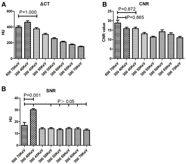
The 40 keV images in the 300 mgI kg−1 group (Group B) had the highest ΔCT value, even though it was not statistically different from that of the 70 keV images in the 600 mgI kg−1 group (Group A) (p > 0.05). The ΔCT value is the increase of the CT value in the renal artery between the contrast-enhanced scan and the plain scan. The higher the ΔCT value, the better the display of renal CTA.
The 40 keV images in Group B had the highest SNR and CNR values, while the SNR was significantly higher than that of the 70 keV images in Group A (p < 0.05), the CNR values were not significantly different between the two image sets (p > 0.05). The comparison of ΔCT value, SNR value, and CNR value between two the groups with different reconstructions are listed in Tables 4 and 5 and Figure 1.
Table 4.
Comparison of ΔCT, SNR and CNR between the images obtained by the different reconstruction methods
| ΔCT | SNR | CNR | Radiologist 1 | Radiologist 2 | |
|---|---|---|---|---|---|
| 600 70 keV | 398.54 ± 73.68 | 16.91 ± 11.12 | 18.78 ± 6.71 | 3.90 ± 0.55 | 4.00 ± 0.65 |
| 300 40 keV | 469.77 ± 86.95 | 30.19 ± 4.41 | 15.52 ± 3.32 | 4.40 ± 0.68 | 4.50 ± 0.60 |
| 300 45 keV | 376.81 ± 72.79 | 13.97 ± 4.61 | 14.86 ± 3.09 | 3.55 ± 0.83 | 3.70 ± 0.80 |
| 300 50 keV | 306.46 ± 59.79 | 14.11 ± 4.43 | 13.09 ± 3.09 | 2.95 ± 0.83 | 3.15 ± 0.88 |
| 300 55 keV | 253.12 ± 49.54 | 13.31 ± 4.27 | 12.20 ± 2.52 | 2.35 ± 0.49 | 2.45 ± 0.61 |
| 300 60 keV | 211.12 ± 38.78 | 14.19 ± 4.40 | 14.18 ± 4.40 | 2.05 ± 0.51 | 2.05 ± 0.69 |
| 300 65 keV | 176.79 ± 32.55 | 13.96 ± 4.57 | 12.83 ± 3.79 | 1.45 ± 0.51 | 1.40 ± 0.50 |
| 300 70 keV | 150.75 ± 29.22 | 12.90 ± 4.18 | 11.00 ± 3.17 | 1.35 ± 0.49 | 1.35 ± 0.49 |
| F Value | 72.67 | 20.56 | 7.54 | 65.87 | 64.85 |
| p-value | 0.000 | 0.000 | 0.000 | 0.000 | 0.000 |
| _____ | _____ | _____ | κ = 0.825 | ||
CNR, contrast-to-noise ratio; SNR, signal-to-noise ratio.
The 40 keV images in Group B had the highest image quality and subjective score.
Table 5.
One-way ANOVA for ΔCT, SNR and CNR between the images obtained by the different reconstruction methods
| Group A (600 mgI kg−1) | Group B (300 mgI kg−1) | ΔCT | SNR | CNR | Radiologist 1 | Radiologist 2 |
|---|---|---|---|---|---|---|
| p-value | ||||||
| 70 keV | 40 keV | 0.409 | 0.001 | 0.865 | 0.312 | 0.096 |
| 45 keV | 1.000 | 0.999 | 0.872 | 0.948 | 0.559 | |
| 50 keV | 0.003 | 1.000 | 0.048 | 0.004 | 0.001 | |
| 55 keV | 0.000 | 0.987 | 0.033 | 0.000 | 0.000 | |
| 60 keV | 0.000 | 1.000 | 0.002 | 0.000 | 0.000 | |
| 65 keV | 0.000 | 0.999 | 0.043 | 0.000 | 0.000 | |
| 70 keV | 0.000 | 0.960 | 0.002 | 0.000 | 0.000 | |
CNR, contrast-to-noise ratio; SNR, signal-to-noise ratio.
The 40 keV images in Group B had the highest SNR and CNR values.
Figure 1.
Comparison of the histogram of ΔCT, SNR and CNR. CNR, contrast-to-noise ratio; SNR, signal-to-noise ratio.
Subjective image evaluation
For the image quality evaluation, the κ value was 0.825 for the two radiologists, indicating a good consistency.
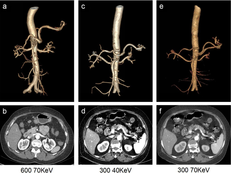
In Group A, both radiologists gave ≥3 points in their subjective evaluations, and the renal artery CTA image quality met the diagnostic needs. In Group B, the subjective scores of the 45 keV images, the 40 keV images and the 50 keV images meet the requirements. There were no significant differences in the subjective scores between the 70 KeV images in Group A and the 45 KeV in Group B (p > 0.05), indicating that the image quality of both images were similar. The image quality of the 40 keV images in Group B was higher than that of the 70 keV images in Group A, and the difference was statistically significant (p < 0.05). Therefore, the 40 keV images in Group B with the lower contrast agent concentration was determined to be the best choice for the subjective evaluation. The subjective scores of renal CTA are listed in Tables 4 and 5. Renal CTA images of different groups are shown in Figure 2.
Figure 2.
(a, b) VR reconstruction of renal CTA with a contrast dose of 600 mgI kg−1 at the energy level of 70 keV in the dual-energy spectral CT imaging, and the corresponding cross-sectional image. (c–f) VR reconstruction of the same patient’s renal CTA with a contrast dose of 300 mgI kg−1 at the energy levels of 40 keV and 70 keV, respectively, and the corresponding cross-sectional images. CTA, CT angiography; VR, volume rendering.
Discussion
Contrast-induced nephropathy (CIN) is the impairment of renal function that cannot be explained by other reasons within 3 days of administration of iodine contrast media. The diagnostic criteria is defined as an increase of ≥25% in serum creatinine (SCr) or an increase of >44 µmol/L in the absolute SCr value within 48–72 h of the i.v. contrast administration.19,20 CIN has become the third most common cause of hospital-acquired acute kidney injury, with an incidence of approximately 11%, especially in the patients with diabetes or pre-existing renal insufficiency.10,11 Currently, no effective measures can be taken to prevent CIN. The European Society of Cardiology recommends solely the use of a hydration therapy or reducing contrast dose to lower the risk of CIN. Therefore, the means to reduce the dose of contrast media, along with ensuring a better image quality has become an issue of concern among the radiologists.
Filtered backprojection (FBP) has been widely used clinically and serves as the “gold-standard” for CT reconstructions for many years.21 However, its weakness in reducing the noise and artifacts at low radiation doses, as well as neglecting the influence of quantum noise and electronic noise on raw data during the acquisition process, seriously limits its applications. The iterative reconstruction technology was first used in the image reconstruction of the positron emission tomography (PET) and single photon emission computed tomography (SPECT). Its primary disadvantage was the slow reconstruction speed. Nevertheless, as the computing power of the computers greatly advanced in recent years, the reconstruction time of iterative reconstruction was significantly shortened, making this technology clinically applicable.
Iterative reconstructions apply different weights to balance noise reduction and spatial resolution loss. In ASIR, different weights in 0–100% are used to adjust the image quality. Theoretically, the higher the ASIR weight, the stronger the noise reduction ability is. However, an overly high ASIR weight may cause the images to be over smoothed, showing “plastic-like artifacts”, which in turn reduces the image quality and thus fails to meet the requirements of correct clinical diagnosis.22 The study by Singh et al showed that appropriate ASIR weights should be chosen under different circumstances. For example, the CTDIvol for abdominal scan was 8.4 mGy, and 30–50% ASIR was an appropriate choice. Therefore, the 40% ASIR was employed for image reconstruction in our study to explore its application in low-contrast CTA of the renal arteries.
Iterative reconstruction algorithms have been widely used in the reconstruction of blood vessels in various parts of the body to reduce the radiation dose. Compared with the traditional FBP, Iterative reconstructions have the advantage of obtaining high-quality reconstructed images under low signal (low dose) conditions. Pontana et al23 used a low-dose examination on 80 patients undergoing follow-up CTA of the chest. The tube current was adjusted to ensure that the CTDI was reduced by 50%. Their results showed that compared with the standard dose FBP images, iterative reconstruction for low-dose CTA had the same subjective image quality and improved objective measurements. Han et al13 selected 80 living kidney donors and used ASIR-V in the low-dose CT angiography of the renal artery. Their results also demonstrated similar subjective image quality and improved objective measurements in ASIR-V low dose CTA compared with the standard CTA with 58% radiation dose reduction. In our study, ASIR was used to reduce image noise of low energy images which have tendency to have higher image noise. Our study illustrated that the ASIR method generated images at the low energy levels with satisfactory quality for diagnosis which were consistent with these previous reports.
The use of low tube voltages and low photon energies (low keV) in renal artery CTA produces higher vascular enhancement which is an effective way to maintain adequate CT number at reduced contrast dose. Almutairi et al24 used only 50% contrast medium volume and demonstrated that images reconstructed at 65 keV with 50% ASIR provided results that were comparable to those with routine contrast medium volume. Patino et al25 used low-energy virtual monochromatic CTA images (at 40 keV and 50 keV) at about 50% of the iodine dose to provide adequate intravascular attenuation and diagnostic quality for aortic assessment. The results in our study showed that the use of 40 keV virtual monochromatic CTA images generate similar enhancement in renal arteries as the conventional CTA with only 50% of the contrast dose, consistent with the above-mentioned studies.
The limitations of this study were that, only an empirical 40% ASIR was employed to reconstruct the images. For CT angiograms, higher percentages of ASIR maybe advantageous. Further study is required to explore the optimal weights for the iterative reconstruction. Due to the limited number of cases, the difference in the detection rate of the renal artery diseases by the two scanning methods was not investigated.
In summary, contrast dose may be reduced by 50% while maintaining image quality by using lower energy images combined with ASIR in renal dual-energy CTA.
REFERENCES
- 1.Rubin GD, Leipsic J, Joseph Schoepf U, Fleischmann D, Napel S. CT angiography after 20 years: a transformation in cardiovascular disease characterization continues to advance. Radiology 2014; 271: 633–52. doi: 10.1148/radiol.14132232 [DOI] [PMC free article] [PubMed] [Google Scholar]
- 2.Pang L, Zhao Y, Dong H, Shi H, Yang W, Zhang H, et al. High-pitch dual-source computed tomography renal angiography comparison with conventional Low-Pitch computed tomography angiography: image quality, contrast medium volume, and radiation dose. J Comput Assist Tomogr 2015; 39: 737–40. doi: 10.1097/RCT.0000000000000268 [DOI] [PubMed] [Google Scholar]
- 3.Ippolito D, Talei Franzesi C, Fior D, Bonaffini PA, Minutolo O, Sironi S. Low kV settings CT angiography (cta) with low dose contrast medium volume protocol in the assessment of thoracic and abdominal aorta disease: a feasibility study. Br J Radiol 2015; 88: 20140140. doi: 10.1259/bjr.20140140 [DOI] [PMC free article] [PubMed] [Google Scholar]
- 4.Ki YJ, Kwon SA, Kim HL, Seo JB, Chung WY. The prevention of contrast induced nephropathy by sarpogrelate: a prospective randomized controlled clinical trial. J Korean Med Sci 2019; 34: e261. doi: 10.3346/jkms.2019.34.e261 [DOI] [PMC free article] [PubMed] [Google Scholar]
- 5.Rudnick MR, Leonberg-Yoo AK, Litt HI, Cohen RM, Hilton S, Reese PP. The controversy of contrast-induced nephropathy with intravenous contrast: what is the risk? Am J Kidney Dis 2020; 75: 105–13. doi: 10.1053/j.ajkd.2019.05.022 [DOI] [PubMed] [Google Scholar]
- 6.Faucon A-L, Bobrie G, Clément O. Nephrotoxicity of iodinated contrast media: from pathophysiology to prevention strategies. Eur J Radiol 2019; 116: 231–41. doi: 10.1016/j.ejrad.2019.03.008 [DOI] [PubMed] [Google Scholar]
- 7.van der Molen AJ, Reimer P, Dekkers IA, Bongartz G, Bellin M-F, Bertolotto M, et al. Post-contrast acute kidney injury - Part 1: Definition, clinical features, incidence, role of contrast medium and risk factors : Recommendations for updated ESUR Contrast Medium Safety Committee guidelines. Eur Radiol 2018; 28: 2845–55. doi: 10.1007/s00330-017-5246-5 [DOI] [PMC free article] [PubMed] [Google Scholar]
- 8.Shah R, Le FK, Labroo A, Khan MR. Contrast-associated acute kidney injury. Quant Imaging Med Surg 2020; 10: 891–4. doi: 10.21037/qims.2020.03.20 [DOI] [PMC free article] [PubMed] [Google Scholar]
- 9.Eng J, Wilson RF, Subramaniam RM, Zhang A, Suarez-Cuervo C, Turban S, et al. Comparative effect of contrast media type on the incidence of contrast-induced nephropathy: a systematic review and meta-analysis. Ann Intern Med 2016; 164: 417–24. doi: 10.7326/M15-1402 [DOI] [PubMed] [Google Scholar]
- 10.Iyama Y, Nakaura T, Iyama A, Kidoh M, Katahira K, Oda S, et al. Feasibility of iterative model reconstruction for unenhanced lumbar CT. Radiology 2017; 284: 153–60. doi: 10.1148/radiol.2017161966 [DOI] [PubMed] [Google Scholar]
- 11.Zhao Y, Zuo Z, Cheng S, Wu Y. CT pulmonary angiography using organ dose modulation with an iterative reconstruction algorithm and 3D smart mA in different body mass indices: image quality and radiation dose. Radiol Med 2018; 123: 676–85. doi: 10.1007/s11547-018-0899-y [DOI] [PubMed] [Google Scholar]
- 12.Sun J, Yang L, Zhou Z, Zhang D, Han W, Zhang Q, et al. Performance evaluation of two iterative reconstruction algorithms, MBIR and ASIR, in low radiation dose and low contrast dose abdominal CT in children. Radiol Med 2020; 125: 918–25. doi: 10.1007/s11547-020-01191-1 [DOI] [PubMed] [Google Scholar]
- 13.Han WK, Na JC, Park SY. Low-dose CT angiography using ASiR-V for potential living renal donors: a prospective analysis of image quality and diagnostic accuracy. Eur Radiol 2020; 30: 798–805. doi: 10.1007/s00330-019-06423-1 [DOI] [PubMed] [Google Scholar]
- 14.Li K, Garrett J, Ge Y, Chen G-H. Statistical model based iterative reconstruction (MBIR) in clinical CT systems. Part II. experimental assessment of spatial resolution performance. Med Phys 2014; 41: 071911. doi: 10.1118/1.4884038 [DOI] [PMC free article] [PubMed] [Google Scholar]
- 15.Ren Z, Zhang X, Hu Z, Li D, Liu Z, Wei D, et al. Application of adaptive statistical iterative Reconstruction-V with combination of 80 kV for reducing radiation dose and improving image quality in renal computed tomography angiography for slim patients. Acad Radiol 2019; 26: e324–32. doi: 10.1016/j.acra.2018.12.021 [DOI] [PubMed] [Google Scholar]
- 16.Prezzi D, Owczarczyk K, Bassett P, Siddique M, Breen DJ, Cook GJR, et al. Adaptive statistical iterative reconstruction (ASIR) affects CT radiomics quantification in primary colorectal cancer. Eur Radiol 2019; 29: 5227–35. doi: 10.1007/s00330-019-06073-3 [DOI] [PMC free article] [PubMed] [Google Scholar]
- 17.Mitsumori LM, Shuman WP, Busey JM, Kolokythas O, Koprowicz KM. Adaptive statistical iterative reconstruction versus filtered back projection in the same patient: 64 channel liver CT image quality and patient radiation dose. Eur Radiol 2012; 22: 138–43. doi: 10.1007/s00330-011-2186-3 [DOI] [PubMed] [Google Scholar]
- 18.Sahani DV, Kalva SP, Hahn PF, Saini S. 16-MDCT angiography in living kidney donors at various tube potentials: impact on image quality and radiation dose. AJR Am J Roentgenol 2007; 188: 115–20. doi: 10.2214/AJR.05.0583 [DOI] [PubMed] [Google Scholar]
- 19.McCullough PA. Contrast-Induced acute kidney injury. J Am Coll Cardiol 2008; 51: 1419–28. doi: 10.1016/j.jacc.2007.12.035 [DOI] [PubMed] [Google Scholar]
- 20.Liang M, Yang S, Fu N. Efficacy of short-term moderate or high-dose rosuvastatin in preventing contrast-induced nephropathy: a meta-analysis of 15 randomized controlled trials. Medicine 2017; 96: e7384. doi: 10.1097/MD.0000000000007384 [DOI] [PMC free article] [PubMed] [Google Scholar]
- 21.Vardhanabhuti V, Ilyas S, Gutteridge C, Freeman SJ, Roobottom CA. Comparison of image quality between filtered back-projection and the adaptive statistical and novel model-based iterative reconstruction techniques in abdominal CT for renal calculi. Insights Imaging 2013; 4: 661–9. doi: 10.1007/s13244-013-0273-5 [DOI] [PMC free article] [PubMed] [Google Scholar]
- 22.Elmokadem AH, Ibrahim EA, Gouda WA, Khalek Abdel Razek AA, Khalek A. Whole-Body computed tomography using low-dose biphasic injection protocol with adaptive statistical iterative reconstruction V: assessment of dose reduction and image quality in trauma patients. J Comput Assist Tomogr 2019; 43: 870–6. doi: 10.1097/RCT.0000000000000907 [DOI] [PubMed] [Google Scholar]
- 23.Pontana F, Pagniez J, Duhamel A, Flohr T, Faivre J-B, Murphy C, et al. Reduced-dose low-voltage chest CT angiography with Sinogram-affirmed iterative reconstruction versus standard-dose filtered back projection. Radiology 2013; 267: 609–18. doi: 10.1148/radiol.12120414 [DOI] [PubMed] [Google Scholar]
- 24.Almutairi A, Sun Z, Poovathumkadavi A, Assar T. Dual energy CT angiography of peripheral arterial disease: feasibility of using lower contrast medium volume. PLoS One 2015; 10: e0139275. doi: 10.1371/journal.pone.0139275 [DOI] [PMC free article] [PubMed] [Google Scholar]
- 25.Patino M, Parakh A, Lo GC, Agrawal M, Kambadakone AR, Oliveira GR, et al. Virtual monochromatic dual-energy aortoiliac CT angiography with reduced iodine dose: a prospective randomized study. AJR Am J Roentgenol 2019; 212: 467–74. doi: 10.2214/AJR.18.19935 [DOI] [PubMed] [Google Scholar]


