
Figure 3. FIGURE 3: Hypoxic treatment aggravates ER stress-induced cellular damage.
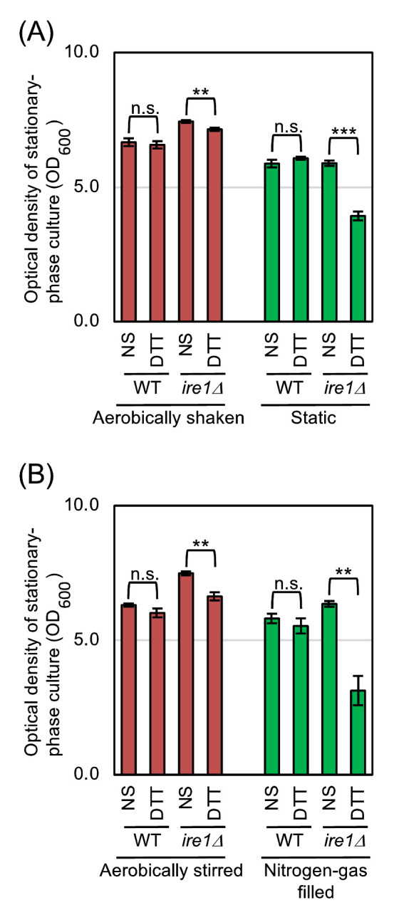
(A) Wild-type BY4742 cells and the congenic ire1Δ mutant were cultured as shown in Fig. 1A, and optical density of the cultures was measured 18 hr after culture start. For DTT treatment, we added DTT (0.5 mM final conc.) into media 4 hr after culture start, and further performed the culturing for 14 hr. (B) The same strains used in panel A were cultured as shown in Fig. 2A, and optical density of the cultures was measured 18 hr after culture start. For DTT treatment, we added DTT (0.5 mM final conc.) into media 4 hr after culture start, and further performed the culturing for 14 hr. n.s. (not significant): p > 0.05, **: p < 0.01, ***: p < 0.001.