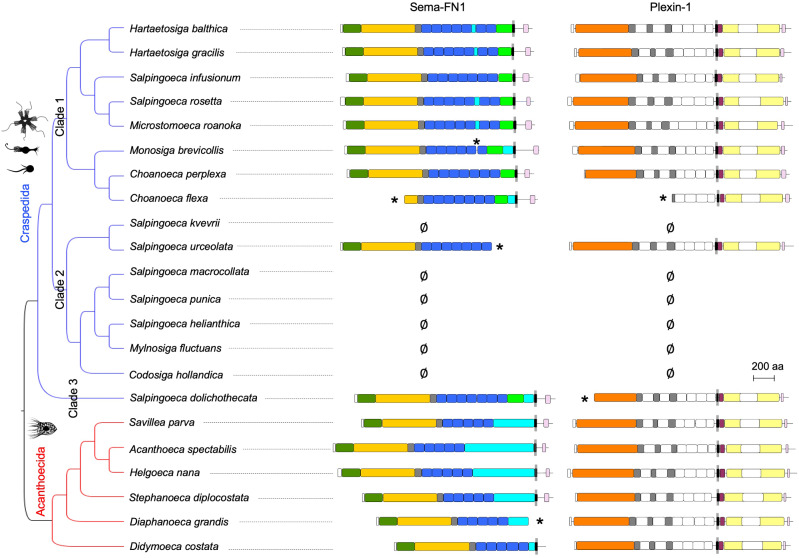
Fig. 2.
Semaphorins and plexins expressed in choanoflagellates. Protein domain structures of semaphorins and plexins, plotted next to the topology of the Bayesian phylogenetic hypothesis of the choanoflagellates analyzed in this study based on 10,816 aligned nucleotides from partial sequences of the genes SSU, LSU, tubA, EFL, and EF-1A (after Carr et al. 2017). Asterisks (*) indicate incomplete sequence and ∅ indicates that the respective semaphorin or plexin mRNA was not detectable. See fig. 1 for colors of domains. See fig. 6 for branch lengths and posterior probabilities of nodes.

