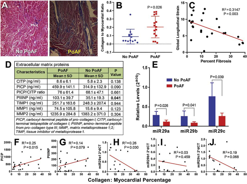
FIGURE 2.
Significant fibrosis in the right atrial appendage of coronary artery bypass graft patients with reduced circulating miR29s and increased procollagen I and III peptide levels in those who developed postoperative atrial fibrillation (PoAF). (A) Representative photographs of Masson’s trichrome staining of right atrial appendage (RAA) histology sections display severe fibrosis (blue) in a PoAF patient (right) compared with a No PoAF patient (left). The individual data points-column scatter graph (B) displays Masson’s trichrome stained collagen (blue) to myocardium (red, total area) ratio, depicting a significantly higher fibrosis (mean±SD and each sample value) in coronary artery bypass graft (CABG) patients who developed PoAF as quantified using ImageJ macro. P ≤ 0.05 was considered significant, n=13 (No PoAF), 15 (PoAF). Linear regression between global longitudinal strain and fibrosis in CABG patients’ RAA histology sections determined by Masson’s trichrome staining and quantified using ImageJ macro to obtain collagen to myocardium ratio (C). Preoperative levels (mean±SD) of markers of collagen and extracellular matrix in patients who developed PoAF versus no-PoAF development after CABG (D). Relative preoperative levels of circulating miR-29a, -b, and -c determined by quantitative polymerase chain reaction were significantly reduced in patients who developed PoAF versus those who remained free of PoAF after CABG during their hospital stay (E). Linear regression between the percentage of right atrial appendage tissue fibrosis and plasma levels of PICP (F), PIIINP (G), and circulating preoperative miR-29a (H), miR-29b (I), and miR-29c (J) levels in coronary artery bypass graft patients.

