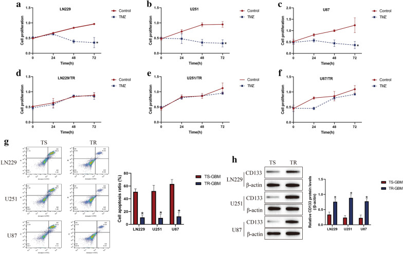
Fig. 1.
Establishment of TR-GBM cells (LN229/TR, U251/TR and U87/TR) by using their parental TS-GBM cells (LN229, U251 and U87). The GBM cells were stimulated by high-dose TMZ (1200 μM) for 0 h, 24 h, 48 h and 72 h, a-f CCK-8 assay was performed to examine cell proliferation abilities. g At 48 h post-treatments, the Annexin V-FITC/PI double staining assay was conducted to measure cell apoptosis in GBM cells. (H) Western Blot analysis was employed to determine the expression levels of CD133 in GBM cells, which were normalized by β-actin. (Note: “TS” indicated "TMZ-sensitive", and “TR” represented “TMZ-resistant”). Each experiment had at least 3 repetitions, and *P < 0.05

