Fig. 2.
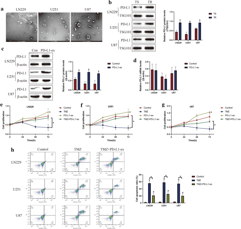
GSCs derived PD-L1-ex increased TMZ-resistance in TS-GBM cells. a The GBM cells derived exosomes were isolated and purified, and the exosomes were observed and photographed by using electron microscope (EM). b The expression levels of PD-L1 in the exosomes were determined by Western Blot analysis, which were normalized by the exosomes marker TSG101. cPD-L1-ex was co-cultured with TS-GBM cells for 24 h, and the expression levels of PD-L1 were examined by performing Western Blot analysis. d Real-Time qPCR was used to examine PD-L1 mRNA levels in TS-GBM cells. e–g CCK-8 assay was performed to measure cell proliferation in TS-GBM cells. h Annexin V-FITC/PI double staining assay was performed to measure cell apoptosis. Each experiment had at least 3 repetitions, and *P < 0.05

