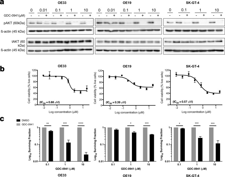
Figure 2.
PI3K inhibition significantly reduces OAC proliferation and survival in vitro. (a) Cells were treated with GDC-0941 or vehicle control for 18 h and tAKT/ pAKT protein levels assessed by Western blot. Western blot is representative of n = 3 repeats. (b) Cells were treated with GDC-0941 at a top concentration of 20 µM and an 8-point dose–response curve generated. The MTS reagent was added to the plates 48 h post-treatment and absorbance measured at 490 nm 4 h later. Cell viability was calculated relative to the corresponding vehicle control for each dose (n = 3). (c) Cells were treated with GDC-0941 or vehicle control and the effect on long-term survival assessed using the clonogenic assay. Data represent the average of n = 3 experimental repeats. Error bars represent mean ± SEM. *p < 0.05, **p < 0.01, ***p < 0.001, ****p < 0.0001. SEM, standard error of the mean.

