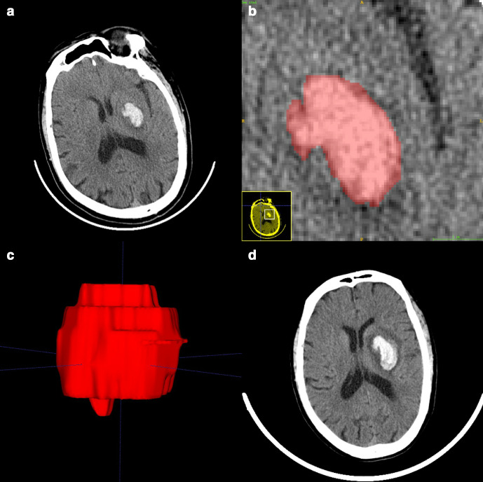
Figure 2.
Manual 3D segmentation of the hematoma. (a) The patient’s baseline CT showed a small hematoma in the left basal ganglia (58 years, male, baseline volume = 7.41 ml, radiomic score = −1.145). (b) Delineation of the lesion using the ITK-SNAP software. (c) Generation of a 3D region of interest. (d) Detection of hematoma expansion on follow-up CT (volume = 16.92 ml, Glasgow outcome scale score = 3). 3D, three-dimensional.

