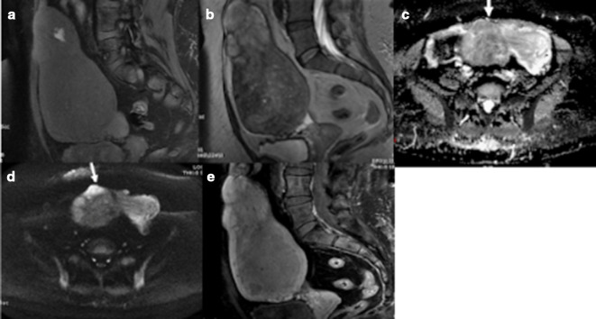
Figure 4.
(a–e) LM with histopathological evidence of haemorrhagic infarction. Intrinsic hyperintensity on fat-suppressed T1WI (a) indicative of haemorrhage; an irregular margin with areas of intrinsic hyperintensity on T2WI (b); restricted diffusion on ADC (c) and DWI (d) and predominantly peripheral enhancement on T1W fat-suppressed post-contrast images (e). ADC, apparentdiffusion coefficient; DWI, diffusion-weighted imaging; LM, leiomyoma; T1WI,T1 weighted image.

