
Figure 5.
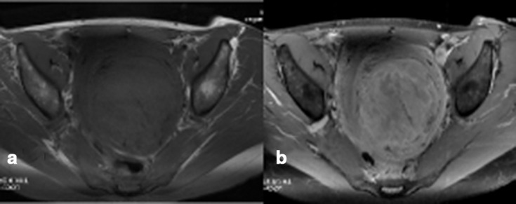
(a, b) Non-infarcted symplastic LM showing predominantly peripheral pattern of contrast enhancement on fat-suppressed T1WI, pre- (a) and post- (b) contrast images, with multiple areas of hypoenhancement in the centre of the lesion. Both radiologists interpreted this as a predominantly peripheral pattern of contrast enhancement but no marginal irregularity/nodularity. LM, leiomyoma;T1WI, T1 weighted image.