
Figure 7.
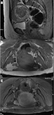
(a–c) LM with no necrosis mentioned in the histopathology report, demonstrating a smooth margin with <50% T2 hyperintensity on T2WI (a), but a predominantly peripheral, “necrotic” pattern of contrast enhancement (7b non-contrast fat suppression T1WI, and 7c post-contrast fat suppression T1WI). LM, leiomyoma;T1WI, T1 weighted image