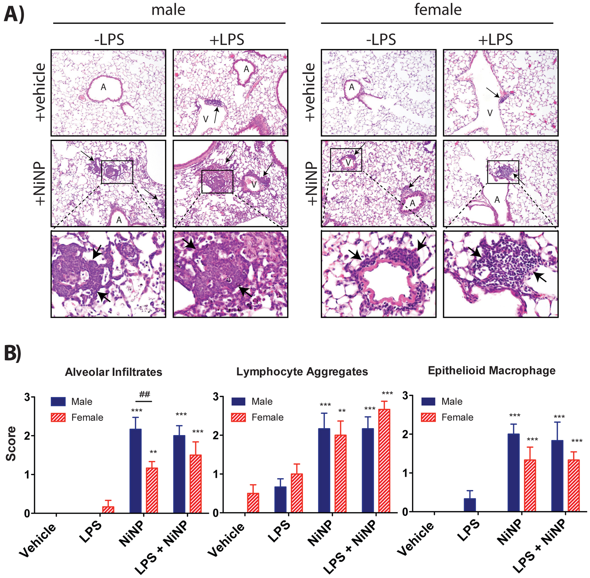
Figure 8.

Lung pathology of male and female mice after repeated subchronic exposure to NiNPs in the absence or presence of LPS. A) Representative hematoxylin and eosin stained lung sections from mice treated with NiNPs or LPS and NiNPs. Inflammatory lesions are indicated by arrows. ‘A’ and ‘V’ indicate airways and vessels, respectively. B) Results of pathology scoring of inflammatory patterns showing relative scores for cellular infiltrates in alveolar lumina, perivascular and peribronchiolar lymphocyte aggregates and dense epithelioid macrophage aggregates. See methods and Supplementary File 1 for scoring system. (n=5–6 mice per group, ***P<0.001 or **P< 0.01 compared to vehicle determined by one-way ANOVA, ##P<0.01 between sexes determined by two-way ANOVA).
