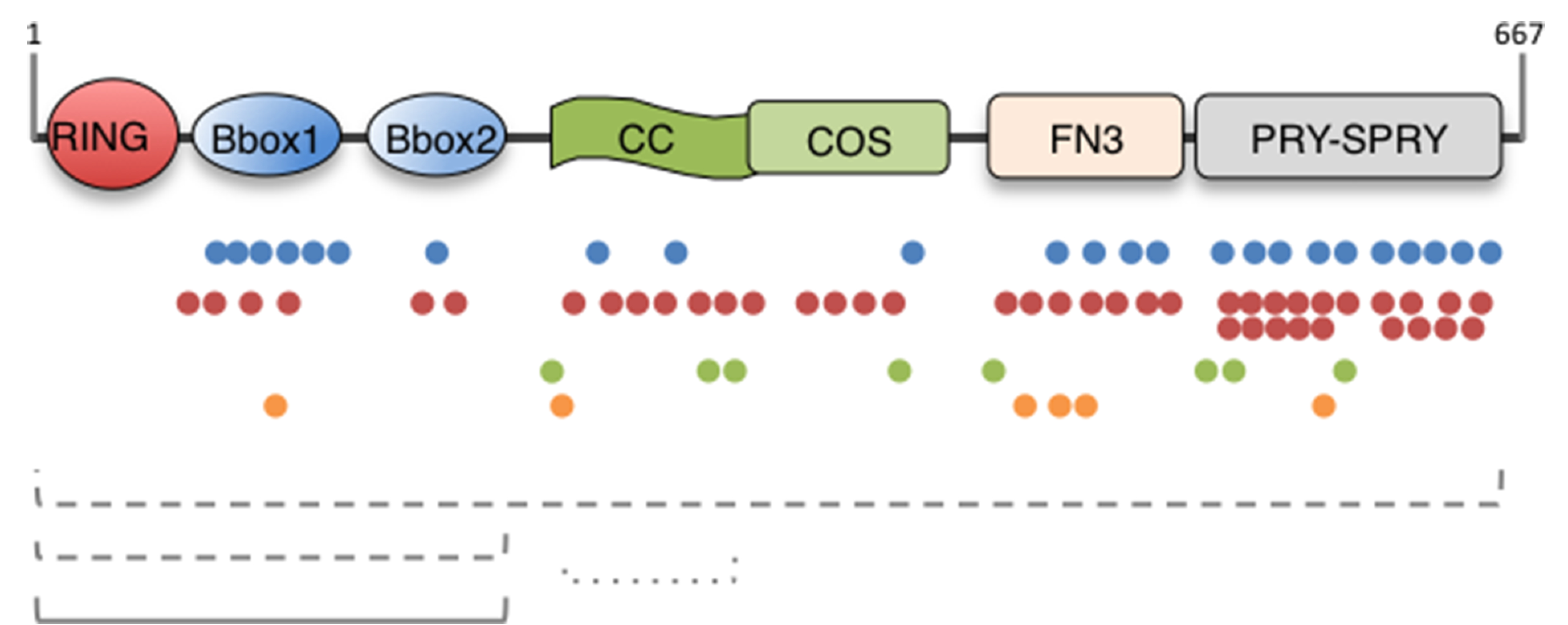
Fig. 1. MID1 protein domain structure and OS-associated mutations.

The domain composition of the MID1/TRIM18 protein is depicted. The MID1 protein is 667 residue-long and the limits of the single domains are following in brackets: RING (10–59), Really Interesting New Gene domain; B-Box (B1, 114–164; B2, 170–212), B-Box domain; CC (219–319), Coiled-coil; COS (320–380), C-terminal subgroup one signature; FN3 (382–472), Fibronectin type III repeat; PRY (483–528), domain associated with SPRY domains; SPRY (538–657), SPla and the RYanodine Receptor. Below the scheme, colour dots represent the different mutations reported so far in OS patients: blue dots, missense mutations; red dots, nonsense and truncating mutations; green dots, splice site mutations; orange dots, inframe indels. The dashed lines represent deletions and rearrangements; the continuous line represents duplications. (For interpretation of the references to colour in this figure legend, the reader is referred to the web version of this article.)
