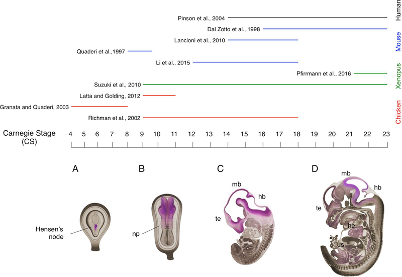
Fig. 4. Distribution of MID1 during embryonic development.
In the upper part, expression of MID1 in specific developmental stages is summarised. The coloured lines indicate the models used in the studies as indicated on the right-hand side. In the bottom part a schematic representation of Mid1 distribution at different stages of embryonic development is shown (pink shading); Carnegie stages (CS) are indicated. A) Mid1 distribution is restricted to the right side of the Hensen’s node; B) the cranial region of the neural plate (np) displays the strongest expression of Mid1 at CS9; C) Mid1 is mainly transcribed in the proliferating compartments of telencephalic vesicle (te), dorsal midbrain (mb) and hindbrain (hb); D) at late embryonic stages, high levels of Mid1 transcript are particularly described in the developing hindbrain and midbrain. Mid1 mRNA is also present in the heart (he) and in several organs of the urogenital system (us). (For interpretation of the references to colour in this figure legend, the reader is referred to the web version of this article.)

