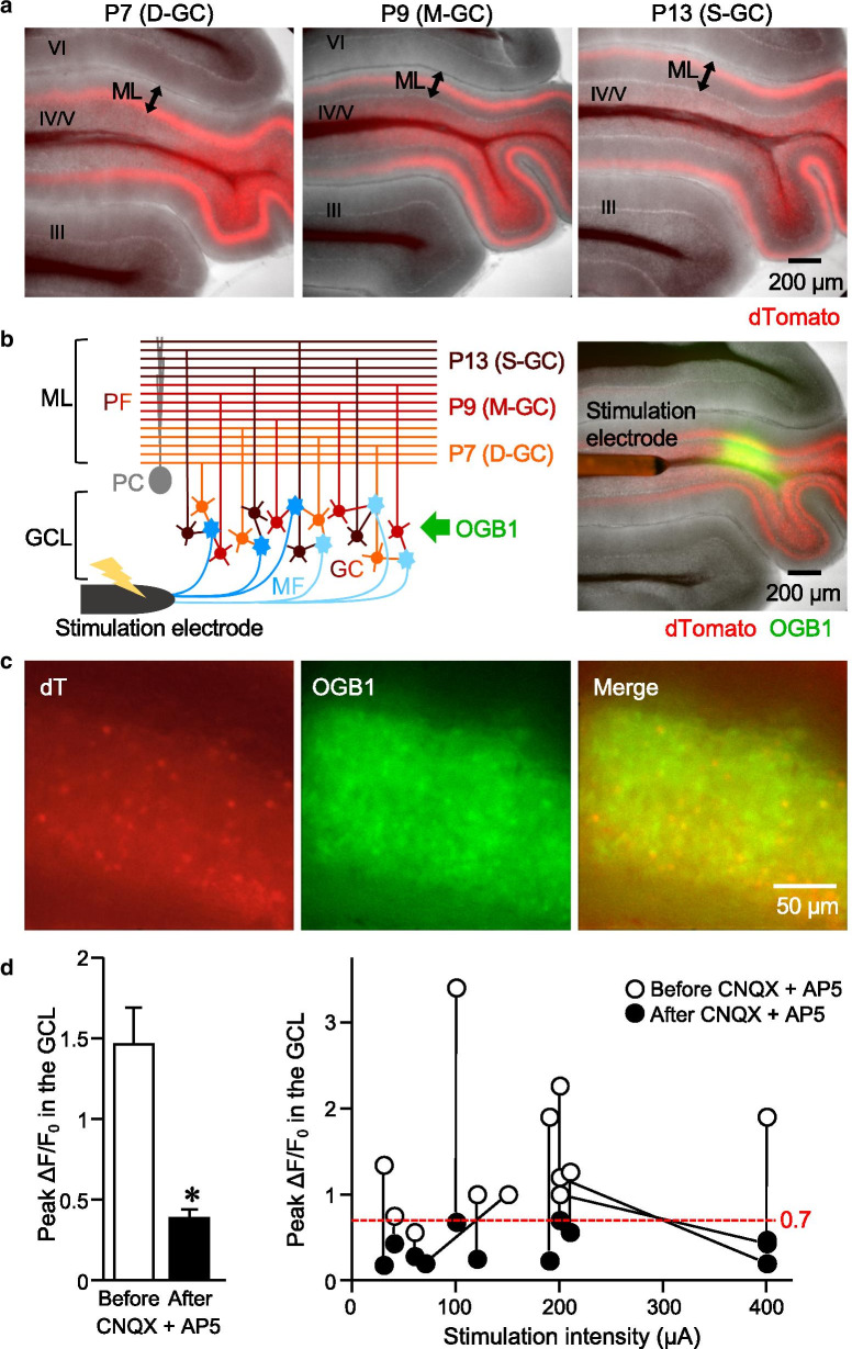
Fig. 1.
Detection of the Ca2+ responses in cerebellar slices labeled with AAV-GABRα6-dT. a Representative images of fresh cerebellar sagittal slices expressing dT (red) in D-GCs (left), M-GCs (middle), and S-GCs (right), which were achieved by the stereotaxic injection of AAV-GABRα6-dT at P7, P9, and P13, respectively. Note that the dT-positive PF bundles are at the deep, middle, and superficial sublayer of the ML. The roman numerals indicate the lobes. b Diagram (left) and an image (right) of experimental setting. OGB1 (green) was loaded in the GCL of cerebellar sagittal slices expressing dT (red), and the bipolar electrode was placed on the white matter to stimulate the MFs. c Magnified images of dT expression and OGB1 loading in GCs of a fresh cerebellar slice. d Peak ΔF/F0 in the whole GCL upon MF stimulation before and after the application of CNQX and AP5. The averaged values (left) and results obtained from individual slices (right) are shown (n = 12 slices, *p = 2.06 × 10⁻⁴, paired sample t-test). Error bars in this and subsequent figures indicate SEM. Exact p values for the datasets in this and subsequent figures are provided in Additional file 1: Table S2

