. 2021 Feb 26;3(2):e200134. doi: 10.1148/rycan.2021200134

Figure:

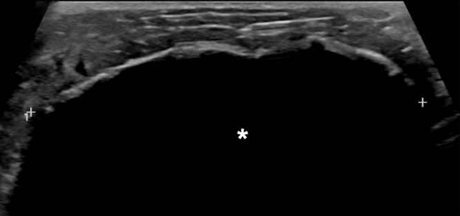
Images in a 73-year-old woman with grade II estrogen receptor/progesterone receptor–positive human epidermal growth factor receptor 2–negative intraductal carcinoma measuring 1.1 cm in size. US images acquired during cryoablation procedure are shown. (a) Long-axis view of tumor (arrow) prior to the procedure. (b) Long-axis view and (c) short-axis view after cryoprobe placement within the tumor (arrow). Arrowhead denotes edge of tumor, and caliper (+) denotes cryoprobe tip. (d) Long-axis view of the ice ball (*) enveloping the tumor. Calipers correspond to the margin of the ice ball.

Images in a 73-year-old woman with grade II estrogen receptor/progesterone receptor–positive human epidermal growth factor receptor 2–negative intraductal carcinoma measuring 1.1 cm in size. US images acquired during cryoablation procedure are shown. (a) Long-axis view of tumor (arrow) prior to the procedure. (b) Long-axis view and (c) short-axis view after cryoprobe placement within the tumor (arrow). Arrowhead denotes edge of tumor, and caliper (+) denotes cryoprobe tip. (d) Long-axis view of the ice ball (*) enveloping the tumor. Calipers correspond to the margin of the ice ball.