. 2021 Mar 26;3(2):e200061. doi: 10.1148/rycan.2021200061

Figure 1:

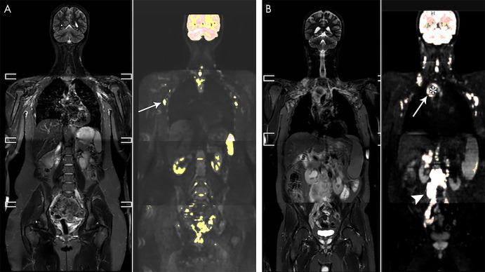
A, Whole-body short-tau inversion-recovery (STIR) image in a 21-year-old female volunteer and overlay of a color-scaled diffusion-weighted image. A region-growing segmentation tool with adaptable threshold allows for the identification of lymph nodes, such as the axillary nodes in this patient (arrow). B, Whole-body STIR image and color-scaled diffusion-weighted image overlay in a 50-year-old man with stage IV mantle cell lymphoma. The large mediastinal mass (arrow) was segmented using a high threshold to include only the most hyperintense component of the mass, excluding the central necrotic portion of the tumor (*). Also shown is a large solid retroperitoneal mass (arrowhead).

A, Whole-body short-tau inversion-recovery (STIR) image in a 21-year-old female volunteer and overlay of a color-scaled diffusion-weighted image. A region-growing segmentation tool with adaptable threshold allows for the identification of lymph nodes, such as the axillary nodes in this patient (arrow). B, Whole-body STIR image and color-scaled diffusion-weighted image overlay in a 50-year-old man with stage IV mantle cell lymphoma. The large mediastinal mass (arrow) was segmented using a high threshold to include only the most hyperintense component of the mass, excluding the central necrotic portion of the tumor (*). Also shown is a large solid retroperitoneal mass (arrowhead).