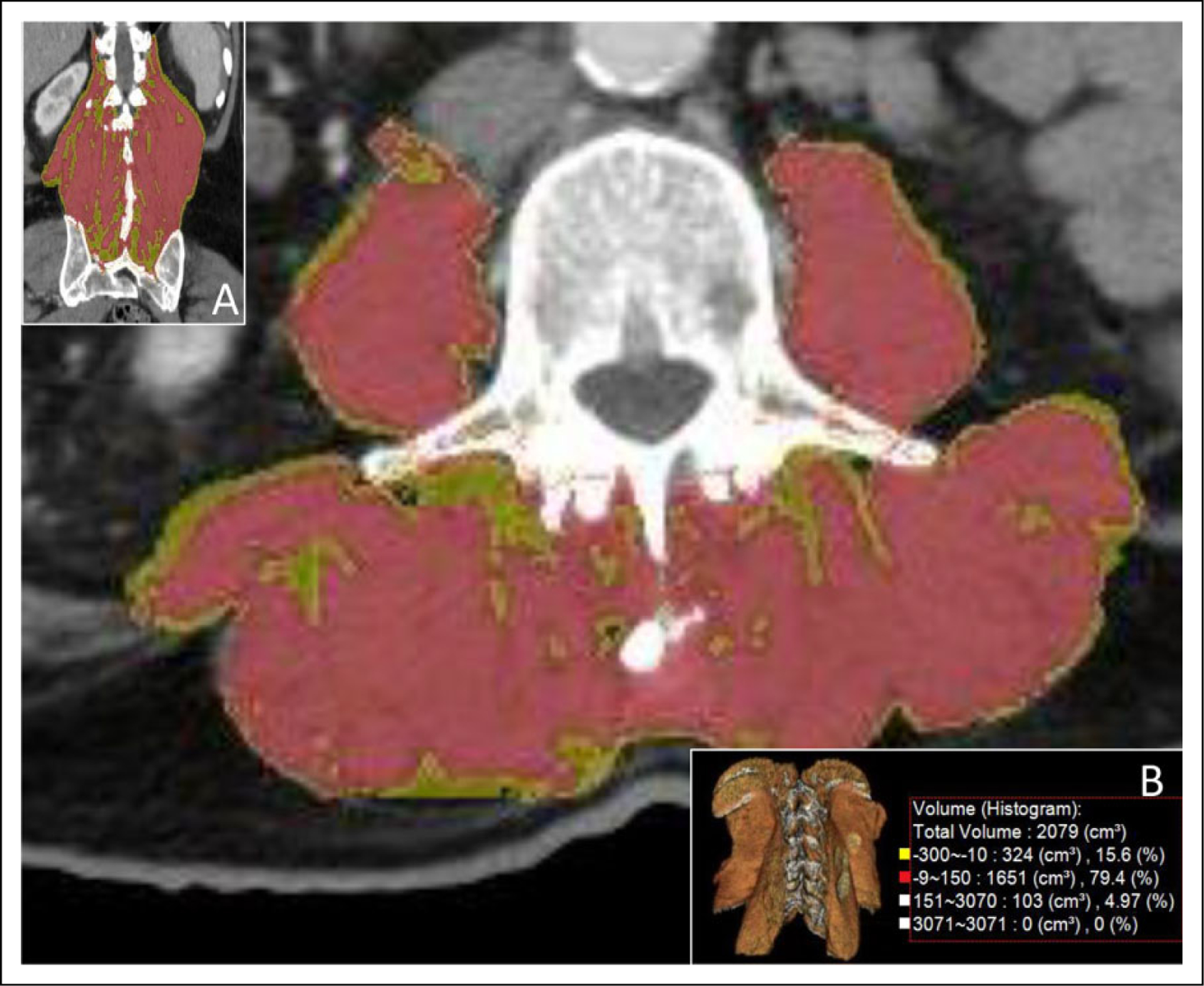
Figure 2.

3D and histogram segmented paraspinal muscles. Semiautomated region-grow tool 3D segmentation of paraspinal muscles with histogram analysis (cm3) using the region grow tool. Parameters including % fat fraction are calculated with fat attenuation range set to −150 to −10 HU and muscle attenuation to −9 to −150 HU. 3D segmentation is shown in axial, coronal, and 3D planes (see insets A and B). Quantitative histogram analysis is shown in inset B. All Paraspinal muscles were segmented from the lower margins of the 12th ribs to the axial slice where the psoas muscle is no longer contiguous with the remaining paraspinal musculature, Segmented muscles included the psoas major anteriorly, quadratus lumborum laterally, the iliocostalis, longissimus, and spinalis posteriorly, and erector spinae posteriorly and inferiorly. 3D indicates 3-dimensional; HU, Hounsfield units.
