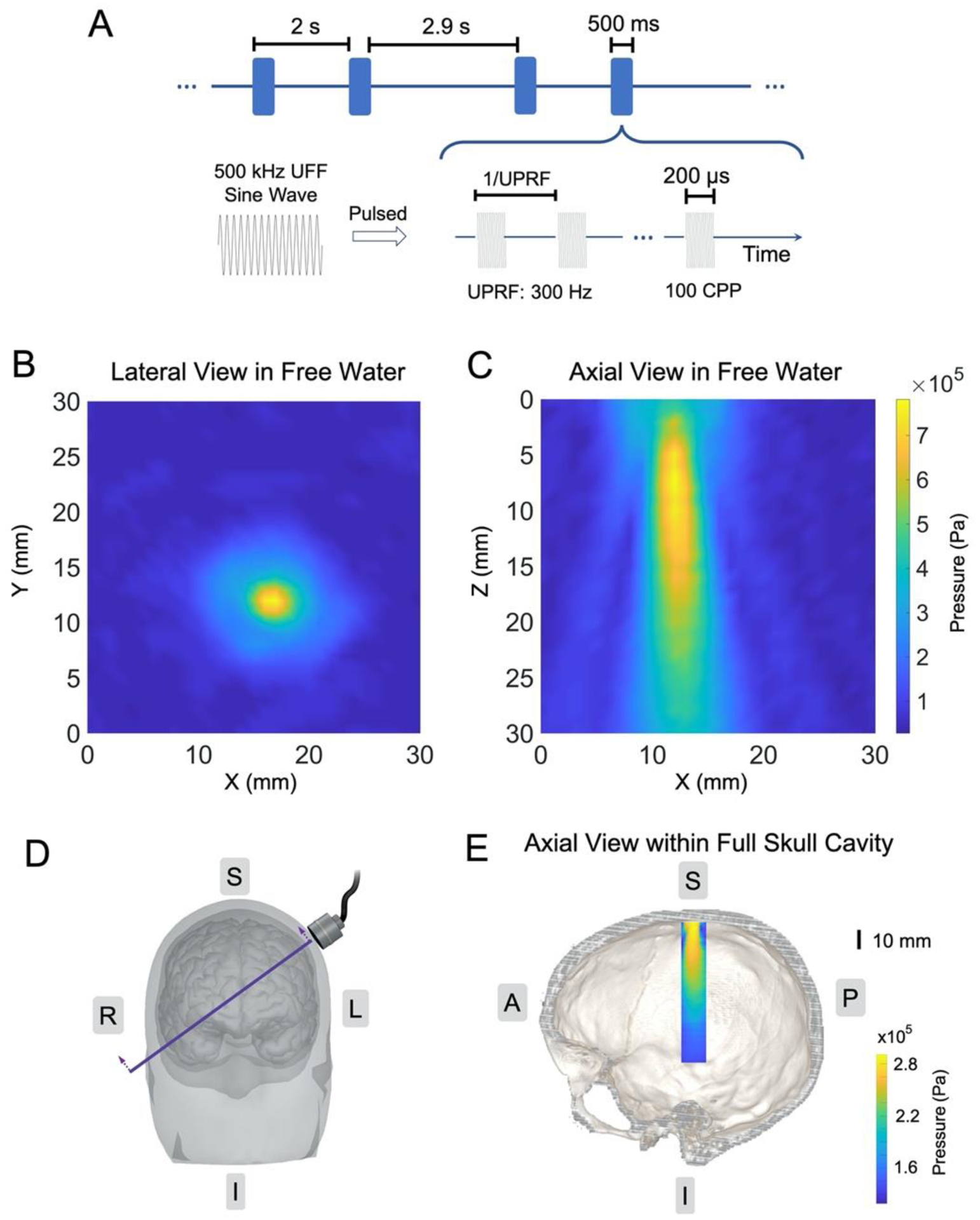
Figure 2.

Ultrasound spatiotemporal profiles. (A) The ultrasound waveform and time sequence. (B-C) Ultrasound pressure field measurements along the lateral direction in free water (B) and along the axial direction in free water (C). (D-E) The illustration of transducer placement over a human head/brain model (D) with the intersectional view of the transcranial ultrasound pressure distribution within a full human skull; the image is co-registered with a human skull model (E).
