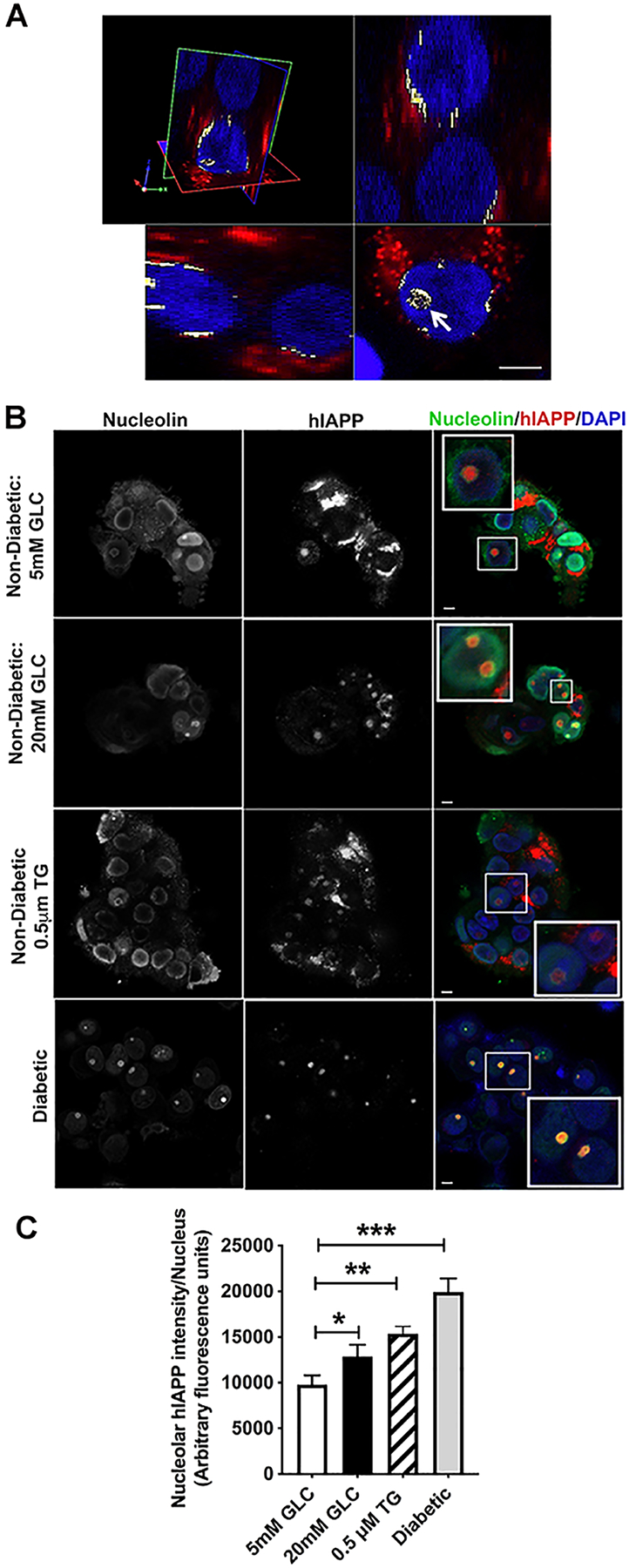
Figure 5. Cellular stress stimulates hIAPP trafficking to the nucleolus.

Non-diabetic human islets were cultured for 4 days in basal (5mM) or high glucose (20mM) media, or with ER stress inducer thapsigargin (0.5μM) for 24 h, and hIAPP trafficking and accumulation examined by immuno-confocal microscopy. (A) 3D immuno-confocal analysis reveals hIAPP-positive puncta in nucleolar and perinuclear regions (white) of cultured human islets. Cells were co-stained with the hIAPP-specific monoclonal antibody (red) and nuclear dye, DAPI (blue). Arrow points to hIAPP-signal within the cell nucleolus. Bar, 5μm. (B) Immuno-confocal analysis of hIAPP and nucleolar marker protein, nucleolin, distributions in control (5mM GLC), ER-stressed (20 mM GLC, 0.5 μM Thapsigargin) and confirmed type-2 diabetic human islets. Cells were co-stained with the hIAPP-specific monoclonal antibody (red), nuclear dye DAPI (blue) and nucleolin specific antibodies (green). Bar, 5μm. (C) Quantitative analysis of the nucleolar hIAPP signal in non-diabetic and T2DM human islets. Significance was established at *p< 0.05, **p< 0.01, ***p< 0.001, ANOVA followed by Tukey’s post hoc comparison test. Data represent mean ± SEM of three independent experiments.
