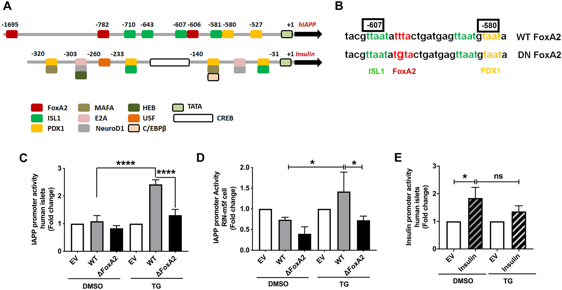
Figure 8. hIAPP promoter activation in ER-stressed pancreatic cells requires a functional FoxA2 binding site.

Freshly isolated human islet or RIN-m5F cells were co-incubated for 24h with renilla (for normalization) and firefly encoding luminescence constructs containing native (WT), mutated FoxA2 (ΔFoxA2) IAPP or WT insulin promoter. Thereafter, transfected cells were incubated with vehicle (DMSO) or 0.5 μm thapsigargin (TG) for an additional 24h. (A) Diagram depicts main transcriptional regulatory sites in hIAPP and insulin promoters. (B) Sequence alignment of WT and DN FoxA2 constructs. Mutated FoxA2 binding site within the hIAPP promoter is shown in red. (C-E) Quantification of insulin and IAPP promoter activity in control (DMSO) and ER stressed (TG) pancreatic cells. Normalized luminescence signal, reflecting promoter activity, is expressed as fold change from empty vector (EV)-transfected islets (set to 1), as described in the method section. Significance was established at *p< 0.05, and ****p< 0.0001, ANOVA followed by Tukey’s post hoc comparison test. Data represent mean ± SEM of three independent experiments.
