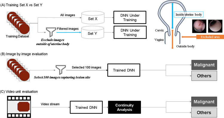
Fig 3.
(A) Schematic of the training method: The training data pertaining to the malignant class were separated into two sets, Set X and Set Y. (B) Schematic of the evaluation method: image by image. (C) Schematic of the evaluation method: video unit. During image-by-image evaluation, 100 images that clearly included the lesion site were extracted from the hysteroscopic video of each patient diagnosed with a malignant tumor (Continuity analysis).

